Preparation and characterization of IRICP
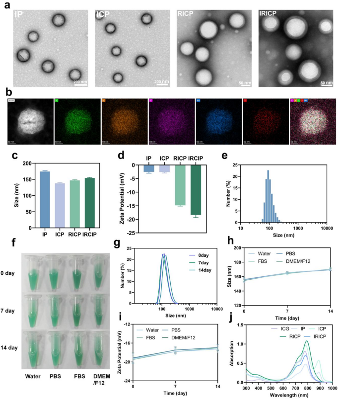
As proven in Scheme 1, ICG and CORM-401 had been emulsified with PLGA to synthesize ICG-CORM-401-PLGA (ICP) cores. Concurrently, the injured RTEC membrane was extracted and combined with ICP at 26 W ultrasound energy. Subsequently, the combination was shaken on a 4℃ shaking desk in a single day to arrange NPs coated with the injured RTEC membrane. With a purpose to exhibit the profitable preparation of IRICP, an analysis of key parameters for IRICP was carried out. The transmission electron microscopy (TEM) photos of IP, ICP, RICP, and IRICP are offered in Fig. 1a. These NPs exhibit a spherical form and exhibit nice dispersion traits. Notably, the outer layer of RICP and IRICP reveals distinct coating when in comparison with IP and ICP counterparts. This statement strongly suggests the profitable encapsulation of the RTEC membrane. As well as, O, N, C, Mn and S components had been detected in ICP by TEM component mapping (Fig. 1b). O, N and C components are current in ICG, PLGA and CORM-401. The S component is present in each ICG and CORM-401. Nevertheless, the component Mn is solely contained in CORM-401. This means the profitable incorporation of CORM-401. As well as, we carried out a comparative evaluation of IP, ICP, RICP, and IRICP and decided their particle measurement and zeta potential utilizing dynamic gentle scattering (DLS). Determine 1c illustrates that the common particle measurement of IP is 174 nm. Apparently, regardless of a rise in package deal contents, the common particle measurement didn’t exhibit a steady increment; as a substitute, it decreased to 137 nm. We postulate that this phenomenon might be attributed to the introduction of CORM-401 which resulted in an enlargement of the solvent system. The typical particle sizes for RICP and IRICP had been measured as 147 nm and 154 nm respectively. Notably, the particle measurement of IRICP was barely bigger than that noticed for RICP. This statement aligns with the findings from TEM depicted in Fig. 1a. The modification of the renal cell membrane in Fig. 1d leads to a major change within the Zeta potential of NPs, shifting from − 2.6 mV to −13.3 mV. Furthermore, when coated with the injured renal cell membrane, the NPs exhibit an additional lower in potential to −18.3 mV. Determine 1e exhibits that the particle measurement distribution of IRICP is especially concentrated within the vary of 68–164 nm. Moreover, to evaluate the steadiness of storage, IRICP was dissolved in numerous solvents together with water, PBS, FBS, and DMEM/F12. The samples had been saved at 4 °C for a period of two weeks. All through the testing interval, IRICP photos captured from completely different solvent circumstances exhibited no vital adjustments (Fig. 1f). Subsequently, the common measurement of IRICP in PBS was measured and Fig. 1g demonstrates that there was minimal alteration in its common measurement over the course of 14 days. Moreover, particle measurement and protein potential measurements had been carried out on IRICP samples immersed in numerous solvents for a span of 14 days (Fig. 1h-i). No notable variations had been noticed throughout this time. These findings spotlight its distinctive long-term storage functionality and stability. As proven in Fig. 1j, The absorption peak of ICG happens at 780 nm. In distinction, the absorption spectra of IP and ICP exhibited substantial alterations, with the wavelength peaks redshifting by roughly 109 nm. Notably, upon encapsulation with the renal cell membrane, the height close to 890 nm vanished. When in comparison with ICG, the RICP peak demonstrated a redshift of roughly 10 nm. Moreover, the absorption peak of IRICP stays centered round 780 nm. The UV-VIS absorption spectra of every materials reveal distinct absorption peaks within the NIR area for every nanomaterial. The outcomes present that the supplies have good NIR optical properties.
Characterization of IRICP. (a) TEM picture of IRICP. (b) Elemental mapping of IRICP. (c) Imply particle measurement of IP, ICP, RICP and IRICP. Imply ± SEM. n = 3. (d) Zeta potentials on the floor of IP, ICP, RICP and IRICP. Imply ± SEM. n = 3. (e) The distribution of particle sizes for IRICP. Common, Imply solely, n = 3. (f) The respective IRICP photos had been preserved in water, PBS, FBS, and DMEM/F12 for durations of seven and 14 days. (g) Alterations within the relative distribution of particle sizes following 7 and 14 days of storage. Imply solely, n = 3. (h) The variation in particle measurement of IRICP was assessed after 7 and 14 days of storage in numerous media. Imply ± SEM. n = 3. (i) The alterations in Zeta potentials following IRICP are assessed after storage in numerous media for 7 and 14 days. Imply ± SEM. n = 3. (j) Absorption spectra within the UV-Vis-NIR vary for IP, ICP, RICP, and IRICP
The multifunctionality of IRICP
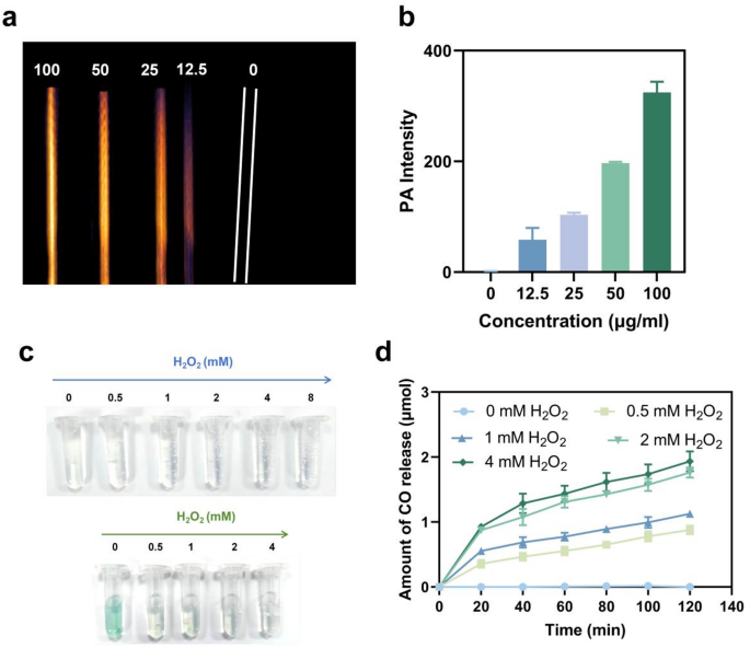
The IRICP concentrations starting from 0 to 100 µg mL−1 had been initially labeled, and their PAI properties at 784 nm had been subsequently measured. The findings demonstrated a linear improve within the PAI sign akin to focus (Fig. 2a-b). The CO launch effectivity of IRICP was additionally assessed. CORM-401 and IRICP had been incubated in PBS with various concentrations of H2O2. As proven in Fig. 2c, after incubation in pure PBS for two h, CORM-401 and IRICP exhibited negligible CO manufacturing. Nevertheless, because the H2O2 focus elevated (0–4 mM), a progressively rising variety of bubbles grew to become distinctly observable within the liquid. Apparently, because the focus of H2O2 will increase, there’s a simultaneous improve within the formation of bubbles within the resolution and a gradual lightening of its inexperienced coloration. This statement additional confirms that IRICP reacts with H2O2 to generate CO gasoline. Subsequently, we employed a real-time CO detector to measure the discharge habits of CO from IRICP. As proven in Fig. 2d, the speed of CO launch was enhanced with rising H2O2 focus, leading to cumulative releases of 1.18 and 1.85 µmol after incubation in 1 and a pair of mM H2O2 for 120 min, respectively.
(a) PAI of IRICP at numerous concentrations assorted from 12.5 to 100 µg mL–1. (b) PA depth of IRICP at various concentrations spanned from 12.5 to 100 µg mL–1. Imply ± SEM. n = 3. (c) Fuel launch profiles of CORM-401 and IRICP below completely different concentrations of H2O2 circumstances. (d) CO launch curves of IRICP below completely different concentrations of H2O2 circumstances. Imply ± SEM. n = 3
In vitro cell-protective actions of IRICP
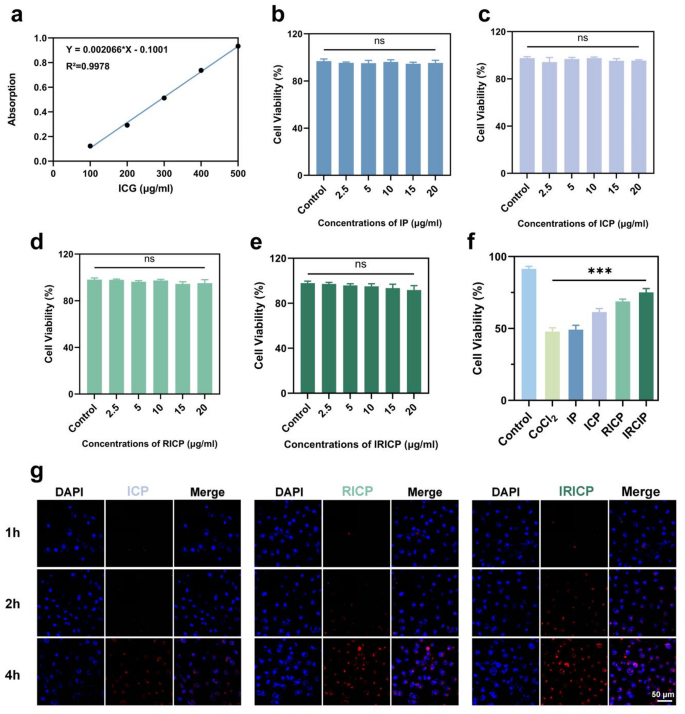
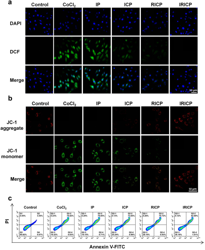
The amount of NPs administered relies on the quantity of encapsulated ICG, which is measured utilizing its normal curve (Fig. 3a). The cytocompatibility and toxicity of every NP (IP, ICP, RICP, IRICP) on HK-2 cells had been subsequently assessed to find out their biocompatibility and potential hostile results (Fig. 3b-e). When HK-2 cells had been uncovered to various concentrations of NPs, the cell viability exhibited a stage akin to the Management group, and any noticed toxicity was negligible. The harm results of various concentrations of CoCl2 on HK-2 cells had been subsequently assessed (Determine S1). Because the focus of CoCl2 elevated, there was a corresponding improve in cell harm, with the cell survival charge reaching roughly 45% at a focus of 700 μm. We subsequently co-incubated numerous NPs with CoCl2-injured cells (Fig. 3f). The therapeutic profit within the IP group was discovered to be minimal in comparison with the CoCl2 group. Within the ICP group, the cell survival charge reached 61.2%. These outcomes demonstrated that ICP successfully launched CO and supplied mobile safety upon addition of CORM-401. Remarkably, each RICP and IRICP teams exhibited enhanced cell safety charges of 66.8% and 75.8%, respectively. Consequently, these findings spotlight the improved therapeutic efficacy conferred by NP coating with renal cell membrane. We subsequently investigated the uptake capability of particular person NPs by HK-2 cells to judge the concentrating on effectivity of injured renal cell membranes (Fig. 3g). Fluorescence photos had been captured at 1, 2, and 4 h. It’s evident from the determine that at 1 and a pair of h, the fluorescence depth was low in each IP and ICP teams, whereas it was increased within the IRICP group. This statement suggests a considerably accelerated mobile uptake charge following coating with injured RTEC membranes. Over time, at 4 h, all teams exhibited NPs ingestion by cells, with the very best fluorescence depth noticed within the IRICP group. These findings point out that encapsulation with injured RTEC membranes can improve mobile uptake effectivity. Oxidative stress serves as a pivotal pathological mechanism within the improvement of AKI. Elevated ranges of oxidative stress amongst sufferers considerably contribute to the development of renal illness. Environment friendly elimination of extreme ROS can successfully safeguard the kidneys towards harm attributable to oxidative stress. With a purpose to additional validate the mobile antioxidant safety of NPs, we employed the DCFH-DA probe for assessing intracellular ROS ranges by measuring cell permeability. As proven in Fig. 4a, in comparison with the Management group, the CoCl2-induced injured cells exhibited a major improve in ROS manufacturing. The fluorescence depth within the IP group was just like that of CoCl2, suggesting an absence of antioxidant impact. Nevertheless, each the ICP and RICP teams demonstrated a discount in ROS manufacturing, with IRICP displaying the weakest DCF fluorescence depth. These findings spotlight the sturdy antioxidant impact of IRICP. Moreover, we utilized mitochondrial membrane potential (ΔΨm) to additional examine the impression of IRICP on oxidative stress-induced damage in renal cells. Typically, a discount in ΔΨm serves as an indicator of early apoptosis, and the JC-1 fluorescent probe was employed to evaluate the ΔΨm throughout all experimental teams. When the ΔΨm is excessive, JC-1 accumulates inside the mitochondrial matrix and emits crimson fluorescence. The alteration in ΔΨm might be detected by observing adjustments in fluorescence coloration. As proven in Fig. 4b, After CoCl2-induced harm to HK-cells, the ΔΨm exhibited a major lower. The depth of inexperienced fluorescence within the IP group was just like that noticed within the CoCl2 group, whereas the ICP, RICP, and IRICP teams displayed a notable discount in inexperienced fluorescence depth. Notably, the IRICP group exhibited the bottom fluorescence depth. These findings recommend that below oxidative stress circumstances, IRICP demonstrates exceptional efficacy in preserving regular membrane potential. As well as, the Annexin V-FITC/PI double staining methodology was employed to evaluate the anti-apoptotic exercise of IRICP. As proven in Fig. 4c, The apoptosis charge within the CoCl2 group and IP group was 79%, whereas the ICP group, RICP group, and IRICP group all exhibited anti-apoptotic results. Amongst them, IRICP demonstrated essentially the most potent anti-apoptotic impact with a lower in apoptosis charge to 62.89%.
(a) Normal curve for ICG. (b–e) Results of various concentrations of IP, ICP, RICP and IRICP on the viability of HK-2 cells. (f) Restoration utilizing numerous supplies following CoCl2-induced cell harm. (g) Cell uptake of ICP, RICP and IRICP. CLSM photos of HK2 cells following co-incubation with numerous NPs for durations of 1, 2, and 4 h. All knowledge represents means ± SEM (n = 3). ***p < 0.001
(a) The ROS fluorescence photos had been in contrast amongst completely different teams after inducing HK-2 cell harm with CoCl2 and subsequent therapy with numerous supplies. (b) Fluorescence photos of JC-1 had been obtained after introducing numerous supplies subsequent to CoCl2-induced cell harm. (c) The HK-2 cells had been subjected to CoCl2-induced harm and subsequently incubated with numerous supplies. The apoptotic course of was assessed utilizing circulation cytometry
Biocompatibility and materials toxicity of IRICP
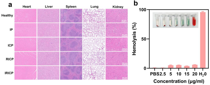
As proven in Fig. 5a, the wholesome mice had been subjected to the injection of varied supplies, and after steady administration for one week, the guts, liver, spleen, lungs, and kidneys had been collected for hematoxylin-eosin (H&E) staining. The HE staining demonstrated that the guts exhibited regular morphology and construction, with no indicators of degeneration or necrosis detected. The liver exhibited diffuse hepatocyte edema, occasional focal necrosis, per delicate lesions. Microscopically, the spleen displayed even distribution of white pulp surrounded by crimson pulp, together with scattered macrophages; total, no vital abnormalities had been famous. Solely native alveolar spacing was barely widened within the lungs, accompanied by dilated and congested blood vessels and minimal inflammatory cell infiltration, indicative of minimal lesions. Gentle tubular dilation with edema was noticed within the kidneys, per minimal lesions. In abstract, the IP, ICP, RICP, and IRICP teams didn’t induce any vital organ harm compared to the wholesome management mice. The findings point out that IRICP exhibits nice biocompatibility and is non-toxic. The biocompatibility and security of IRICP with completely different concentrations had been additional assessed by hemolysis experiments concurrently (Determine. 5b). After putting the IRICP suspension for 3 h, the combination was centrifuged and the hemoglobin content material within the supernatant was measured. We employed phosphate-buffered saline (PBS) because the destructive management, with a hemolysis charge of 0.64%, which is near the perfect 0% hemolysis charge, and deionized water because the constructive management, with a hemolysis charge of 96.86%. These benchmarks had been used to judge the hemolytic efficiency of IRICP inside the focus vary of two.5–20 µg/mL. The outcomes demonstrated that the hemolysis charge of IRICP (0.76–5.97%) was considerably decrease than that of the constructive management, thereby confirming its good blood compatibility.
NIR and PA prognosis of AKI with IRICP
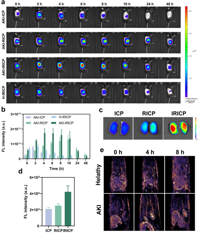
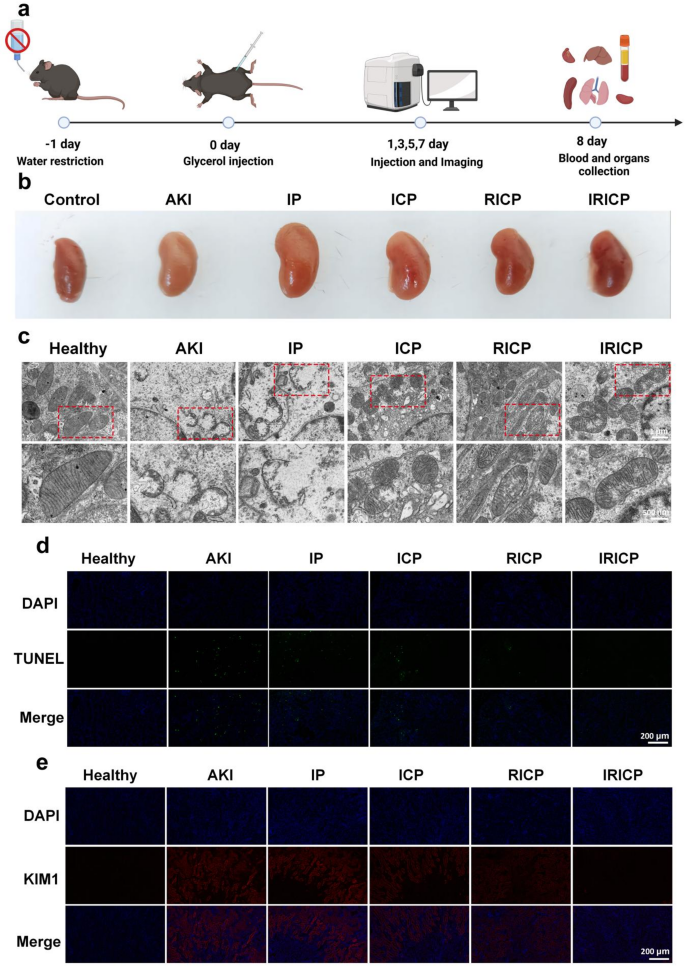
We established AKI mannequin by intramuscular injection of glycerol. The mice had been disadvantaged of water for twenty-four h previous to injection. Subsequently, a 50% glycerol resolution was injected into the leg muscle groups of the mice (Fig. 7a). We then administered numerous NPs to the mice, with the amount of NPs decided primarily based on the quantity of ICG (Fig. 3a). The in vivo distribution of ICG (500 µg mL−1 of ICG, 60µL) was subsequently assessed utilizing an in vivo fluorescence imaging system. A number of angles had been captured throughout imaging classes for the mice (Determine S2), revealing clear accumulation of ICG within the kidney. To facilitate handy statement of ICG distribution inside the physique, we finally chosen the best facet for visualization functions. As proven in Fig. 6a, distinct nanomaterials had been injected into mice, and NIR fluorescence photos had been captured at 0, 2, 4, 6, 8, 10, 24, and 48 h post-administration. In AKI mice, the ICP group confirmed renal accumulation because of elevated vascular permeability after kidney damage, enabling passive concentrating on of nanomaterials to the kidney. The RICP group exhibited a stronger kidney fluorescence sign than the ICP group, indicating enhanced concentrating on by tubular epithelial cell membranes on the damage website. Notably, the IRICP group displayed considerably increased fluorescence depth than the RICP group, highlighting its superior homologous concentrating on skill for injured RTEC membranes. Whereas fluorescence depth weakened within the ICP group after 4 h, each RICP and IRICP teams confirmed rising depth at 4 and 6 h. The IRICP group maintained sturdy fluorescence from 4 to 10 h and even at 48 h, suggesting that injured RTEC membranes alter NP metabolism in AKI mice by enhancing kidney concentrating on and retention. Determine 6b illustrates normalized fluorescence sign intensities at every time level. The mice had been euthanized and renal imaging was carried out 6 h post-injection. Ex vivo imaging (Fig. 6c) and corresponding fluorescence depth quantification (Fig. 6d) had been per the outcomes proven in Fig. 6a, indicating nice renal concentrating on properties of NPs modified by injured RTEC membranes. To evaluate the diagnostic efficacy of IRICP in AKI, we administered an equal dose of IRICP to each the AKI group and the Wholesome group. Subsequently, NIR fluorescence imaging was carried out over a 48-hour interval (Fig. 6a). Compared to the Wholesome group, the AKI group exhibited considerably stronger fluorescence depth all through your complete period. Particularly, whereas the fluorescence depth regularly decreased in wholesome mice ranging from 2 h, it continued to extend in AKI mice. Apparently, the fluorescence depth of the IRICP group peaked at 4 h, exhibiting a major disparity in comparison with that of the wholesome group. Conversely, the RICP group didn’t attain its peak at 4 h and displayed minimal divergence from the wholesome group. The height fluorescence depth within the RICP group was solely achieved after 10 h. This statement means that injured RTEC membranes possess an enhanced capability for speedy accumulation relative to regular RTEC membranes, facilitating expedited and extra pronounced prognosis of AKI. Subsequently, leveraging the fabric properties outlined in our prior findings (Fig. 2a), IRICP demonstrates environment friendly absorption of laser vitality and its conversion into thermal vitality, thereby producing a strong ultrasonic sign through thermoelastic enlargement. Consequently, we assessed the diagnostic functionality of IRICP utilizing PAI. As depicted in Fig. 6e, PA imaging was carried out at 0, 4, and eight h post-injection of IRICP. The outcomes point out that the depth of the photoacoustic sign generated by IRICP below NIR excitation is markedly increased than that of background tissues, sustaining excessive decision even in deep tissues, thus rendering it appropriate for in vivo imaging purposes. At 4 and eight h post-injection, distinct PA alerts had been clearly noticed within the renal area of the AKI group, whereas no vital PA alerts had been detected within the Wholesome group. By means of photoacoustic imaging, IRICP considerably expedites the prognosis of AKI in comparison with standard scientific measurements of Crea and BUN over a 48-hour interval. Furthermore, the spatial distribution and depth of photoacoustic alerts not solely facilitate early prognosis of AKI but additionally allow signal-guided, region-specific remedy, permitting IRICP to launch CORM-401 within the kidneys and subsequently launch CO. This outstanding PA sign could also be attributed to the homologous concentrating on impact of the broken renal cell membrane, which extends the circulation time of IRICP and consequently enhances its accumulation within the kidneys. Moreover, a number of imaging experiments demonstrated that IRICP doesn’t exhibit photobleaching or photothermal degradation below three laser pulse irradiations, indicating its suitability for long-term dynamic monitoring. These experimental findings collectively recommend that, whether or not primarily based on NIR fluorescence imaging or PA imaging, IRICP possesses the potential to diagnose AKI and might monitor therapy responses in actual time through dynamic sign adjustments, thereby offering a basis for customized prognosis and therapy.
(a) In vivo NIR fluorescence photos of various supplies injected into AKI and wholesome mice at numerous time factors. (b) FL sign depth within the kidneys noticed in vivo. Imply ± SEM (n = 3). (c) Kidney was imaged utilizing NIR fluorescence within the ICP, RICP, and IRICP teams. (d) FL sign depth within the kidneys measured ex vivo. Imply ± SEM (n = 3). (e) In vivo PA imaging of AKI mice at 0, 4 and eight h after injection of IRICP (kidney websites are circled)
Remedy of AKI with IRICP
We utilized a glycerin-induced mannequin of AKI to additional validate the therapeutic efficacy of IRICP. AKI mice had been subjected to a number of injections of IRICP at particular time factors, particularly 1, 3, 5, and seven days. On day 8, the mice had been euthanized and serum and tissue samples had been collected for subsequent evaluation, as depicted in Fig. 7b. The kidneys of the AKI group and wholesome group exhibited no discernible variations in look; nevertheless, their coloration appeared pale slightly than rosy, indicative of ischemia. Equally, the kidneys of the IP group and AKI group displayed comparable appearances with a pale coloration per ischemia. Consequently, these findings recommend that the IP intervention didn’t yield any therapeutic results. Notably, the kidneys of the ICP group, RICP group, and IRICP group regularly regained their redness over time, with these from the IRICP group resembling these from the wholesome group to an awesome extent. These findings recommend that IRICP reveals superior therapeutic efficacy. The mitochondria, as essential organelles concerned within the era of ROS inside cells, exhibit morphological alterations that function early indicators of mitochondrial dysfunction attributable to oxidative stress [47]. The renal sections from every experimental group had been examined utilizing TEM to look at the morphology of mitochondria. TEM photos (Fig. 7c) present vital diffuse mitochondrial harm within the proximal renal tubular cells of glycerin-induced AKI mice, marked by wrinkling of the mitochondrial membrane, a lighter stroma, and disruption and fragmentation of ridges, with some being damaged and even lacking. In distinction, the mitochondrial morphology and construction of RTECs within the therapy group confirmed vital enchancment. The mitochondria within the IRICP group exhibited minimal harm with well-preserved ridge constructions and a morphology most just like that noticed within the wholesome group. In superior AKI, intensive apoptosis of the proximal tubules leads to nephron loss and finally accelerates the onset and development of renal failure. TUNEL staining was employed to evaluate whether or not IRICP might mitigate the apoptosis of renal tubular cells in AKI mice (Fig. 7d). The fluorescence alerts had been most pronounced within the AKI group and IP group. The inexperienced fluorescence regularly diminished within the ICP group, RICP group, and IRICP group. Notably, IRICP exhibited the weakest inexperienced fluorescence sign, suggesting its potent anti-apoptotic functionality. The cell membrane glycoprotein Kim-1, a sort I protein, is usually undetectable in wholesome kidneys however turns into detectable in instances of kidney harm. Consequently, Kim-1 serves as a particular biomarker for assessing kidney damage sensitivity and has obtained FDA approval for this goal [48]. Determine 7e exhibits that Kim-1 expression was most pronounced within the AKI and IP teams, primarily discovered at tubular areas, indicating vital harm to tubular cells. In distinction, the IRICP group exhibited a relative fluorescence depth of Kim-1 that decreased to ranges just like these seen within the wholesome group. This means that CO gasoline can successfully scale back ROS on the damage website, thus assuaging hurt to renal tubule cells.
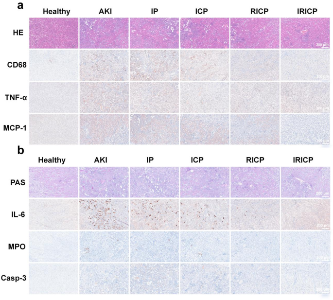
We carried out additional evaluation on every group of H&E-stained kidney sections (Fig. 8a). The renal interstitium, glomeruli, and tubules exhibited regular morphology within the wholesome group, whereas reasonable tubule atrophy, delicate tubule dilation, and reasonable edema had been extra prevalent within the AKI group and IP group. Often, punctate necrosis of tubular epithelial cells, fragments of cell necrosis, and neutrophil infiltration had been noticed inside the lumen. The diploma of kidney damage decreased successively within the ICP group, RICP group, and IRICP group; nevertheless, the therapy impact was most pronounced within the IRICP group with a mobile construction intently resembling normalcy. As AKI progresses, macrophages transfer to the broken kidney, leading to structural hurt and the discharge of inflammatory elements. This course of worsens irritation and rapidly deteriorates renal perform. Determine 8a illustrates that the IP group had the very best macrophage depend, whereas their numbers had been decrease within the ICP and RICP teams. Notably, the IRICP group displayed a pronounced improve in macrophage infiltration. Concurrently, macrophages can secrete pro-inflammatory cytokines comparable to IL-6 and TNF-α, thereby selling irritation and tissue harm. As depicted in Fig. 8a-b, The degrees of TNF-α and IL-6 had been discovered to be considerably elevated within the AKI and IP teams. Conversely, the IRICP group exhibited notably decrease cytokine ranges, indicating a pronounced anti-inflammatory impact of IRICP. Within the pathogenesis of AKI, each immune and renal cells straight generate pro-inflammatory cytokines and chemokines to set off the inflammatory response. Monocyte chemoattractant protein-1 (MCP-1) performs an important function on this response by activating monocytes/macrophages by chemotaxis. As depicted in Fig. 8a, the IRICP group demonstrated markedly lowered MCP-1 ranges compared to the AKI and IP teams. In comparison with HE staining, PAS staining allows a clearer visualization of the glomerular basement membrane. As depicted in Fig. 8b, each AKI and IP teams exhibited slight tubular basement membrane thickening, whereas just a few tubular basement membranes confirmed marginal thickening within the IRICP group. Myeloperoxidase (MPO) serves as an indicator of neutrophil activation, making it a reliable biomarker for evaluating inflammatory standing [49]. As depicted in Fig. 8b, the IRICP group confirmed the bottom ranges of MPO when in comparison with each the AKI and IP teams. Caspase-3 ranges had been measured to judge apoptosis in renal tissue (Fig. 8b). The method of apoptosis is initiated on the molecular stage by proteolytic enzymes belonging to the Caspase household, that are labeled as cysteine proteases. Following renal damage, there was a major upregulation within the expression stage of Caspase-3. Nevertheless, this improve in Caspase-3 expression was counteracted in AKI mice handled with IRICP.
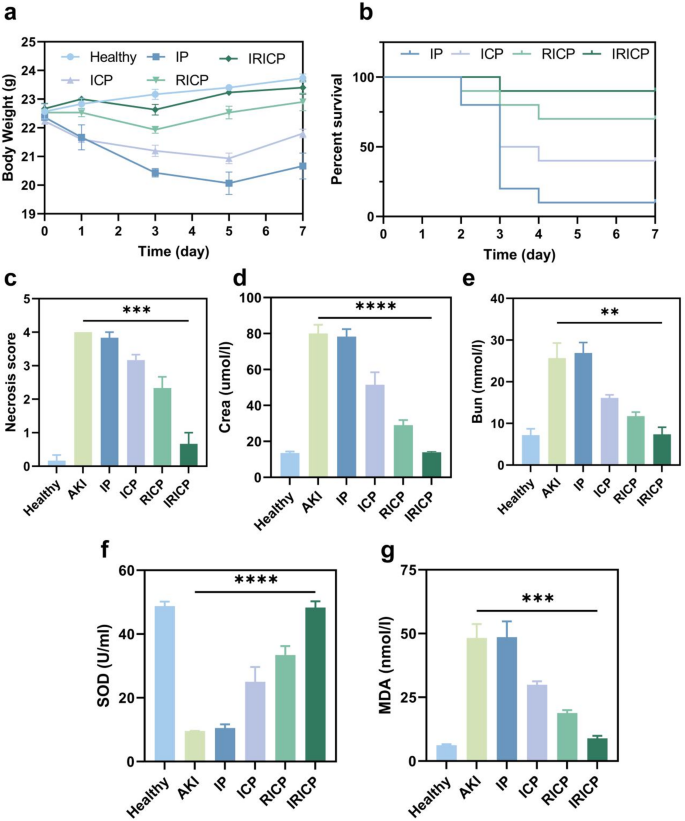
Through the therapy interval, we additionally intently monitored the fluctuations in physique weight of the mice (Fig. 9a). The transient weight achieve (0.2 g) noticed within the IRICP group on the primary day might point out that its early protecting impact delayed metabolic consumption related to AKI. Moreover, the lower in weight after the third day was considerably decrease in comparison with that within the IP group, which additional suggests the inhibitory impact of IRICP on the development of AKI. By the third day, all mice apart from the Wholesome group exhibited weight reduction. The RICP and IRICP teams commenced weight regain on the fourth day. All through the therapy interval, the IP group demonstrated the very best diploma of weight reduction, adopted by the ICP and RICP teams, whereas the IRICP group’s weight remained akin to that of the wholesome group on day 7. Moreover, we generated a survival curve (Fig. 9b), which clearly demonstrated that AKI mice handled with IRICP skilled a major enchancment of their survival charge. Moreover, we assessed and scored the extent of kidney damage inside every experimental group (Fig. 9c). The elevation of Crea and BUN serves as a dependable scientific indicator for assessing renal perform impairment. As depicted in Fig. 9d-e, the IP group exhibited considerably elevated ranges of (Crea) and BUN. Compared to the IP group, each the ICP and RICP teams demonstrated a discount in these parameters amongst AKI mice, with the IRICP group exhibiting essentially the most substantial lower. These outcomes point out that IRICP considerably improves renal perform in mice with AKI. Malondialdehyde (MDA) is the last word byproduct of lipid peroxidation, serving as an oblique indicator of ROS presence and kidney harm. Superoxide dismutase (SOD) is essential within the antioxidant protection mechanism, because it inhibits the lipid peroxidation chain response triggered by free radicals, thus defending cells and tissues from harm attributable to ROS. Consequently, oxidative stress was assessed by measurements of SOD exercise and MDA ranges post-treatment. As depicted in Fig. 9f-g, each ICP and RICP teams exhibited elevated SOD exercise alongside lowered MDA content material; nevertheless, IRICP demonstrated a extra pronounced therapeutic impact. In conclusion, these findings point out that IRICP successfully inhibits irritation, exerts an anti-oxidative stress impact, and reduces apoptosis of renal tubule cells, thereby contributing to kidney restoration.
(a) Diagram illustrating the creation of the AKI mannequin. (b) Photographic photos depicting numerous teams of stable kidneys. (c) Consultant TEM photos of renal sectioned mitochondria (Crimson space represents the magnified location). (d) TUNEL assay was carried out to evaluate apoptosis within the completely different cell teams. (e) Confocal microscopy picture showcasing KIM-1 renal tubules
(a) The intergroup disparities in murine physique weight. (b) Survival curve of mice in numerous therapy teams. (c) Renal damage scoring primarily based on H&E staining outcomes. Serum ranges of (d) Crea, (e) BUN, (f) SOD and (g) MDA in every group. All knowledge represents means ± SEM (n = 3). **p < 0.01, ***p < 0.001, ****p < 0.0001


