Synthesis and characterization of the CR NPs
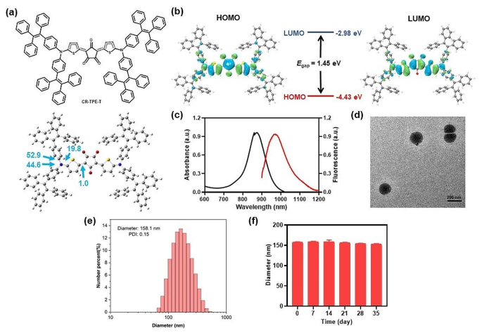
First, the D–π–A–π–D structured CR-TPE-T was synthesized by means of a single-step condensation between croconic acid and N, N-bis(4-(1,2,2-triphenylvinyl) phenyl) thiophen-2-amine in 1:2 equal primarily based on the earlier reviews [32] (Scheme S1). The molecular geometry and digital distribution of CR-TPE-T had been studied by density purposeful concept (DFT) calculations. As proven in Fig. 1a, the optimized ground-state (S0) geometry of CR-TPE-T confirmed an axisymmetric configuration in house; the 2 dihedral angles between TPE and its neighboring thiophene ring had been 52.9° and 44.6°. The dihedral angle between the thiophene ring and the croconic ring was 1.0°, and the entire molecule tended to look as a planar conformation alongside the spine. In CR-TPE-T, the electron densities of the very best occupied molecular orbital (HOMO) had been effectively distributed on the tetraphenylethylene (TPE) donors alongside its conjugated thiophene items, whereas the bottom unoccupied molecular orbital (LUMO) was extra delocalized on the electron-deficient croconaine core, revealing environment friendly intramolecular cost switch inside the molecule. The HOMO and LUMO vitality bandgap of CR-TPE-T was roughly 1.45 eV (Fig. 1b).
Characterization and properties of the CR NPs. (a) Chemical construction and optimized molecular geometry of CR-TPE-T. (b) The calculated frontier molecular orbitals of CR-TPE-T calculated by the density purposeful concept (DFT) calculation technique on the B3LYP/6-31G stage. (c) Absorption and fluorescent spectra (excited wavelength 808 nm) of croconaine dye nanoparticles (CR NPs) in aqueous resolution. (d) Consultant transmission electron microscopy (TEM) picture and (e) Hydrodynamic measurement distribution of CR NPs. scale bar: 200 nm. Polydispersity index (PDI) = 0.15. (f) Dimension of CR NPs after 5 weeks of storage at nighttime at room temperature
For in vivo purposes, the hydrophobic CR-TPE-T was then fabricated into nanoparticles (NPs) (CR NPs) by means of nanoprecipitation utilizing DSPE-mPEG2000 because the encapsulation matrix. In keeping with the normalized absorption spectrophotometer evaluation, the calculated encapsulation effectivity of the CR NPs was 89% (Determine S1). The UV-vis-NIR absorption spectra evaluation of the CR NPs displayed an intense attribute absorption peak of 870 nm and lengthening over 1000 nm, matching very effectively with the organic window (700–1000 nm), which means that it has potential to function a PTA. As well as, the CR NPs confirmed the corresponding fluorescence emission protecting the NIR-II area, which extends to 1200 nm, with most peak at 970 nm, the fluorescence quantum yield of CR NPs in water was calculated to be 0.47% through the use of IR 1061 as a reference (Determine S2), indicating the potential for NIR-II FLI (Fig. 1c).
The TEM picture and DLS leads to Fig. 1d and e indicated a dispersed spherical form of CR NPs with common diameter of ∼ 158 nm, indicating that the NPs would possibly obtain passive tumor accumulation by means of the EPR impact. Moreover, the CR NPs zeta potential was − 33.7 ± 1.71 mV, which favors good stability in a physiological setting (Determine S3). The colloidal stability of CR NPs was additional studied by recording their particle measurement at room temperature for five weeks. Notably, negligible variation of the diameter distribution was noticed for the NPs resolution after storage at room temperature for five weeks, implying good colloidal stability of the CR NPs (Fig. 1f). These outcomes reveal that CR NPs are a promising candidate for NIR-II FLI-guided PTT purposes.
In vitro NIR-II FLI and PTT properties of CR NPs
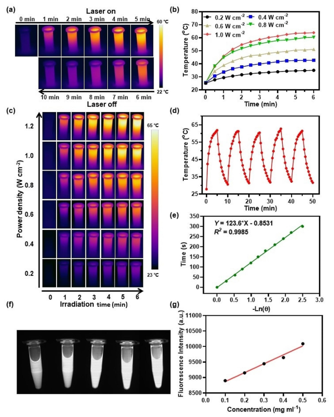
Due to the inherent NIR absorption capability of the CR NPs, we then investigated the PTT conduct of CR NPs in vitro. As proven within the infrared pictures (Fig. 2a) recorded by a thermal digital camera, the CR NPs exhibited an obvious photothermal impact on 808-nm laser irradiation. The laser energy density results on the temperature modifications of CR NPs had been then investigated. It was evident that the profiles of temperature increments of the CR NPs had been positively associated to laser energy density (Fig. 2b). The temperature elevated to roughly 62.5 °C on the energy density of 1 W cm− 2 on 808-nm laser irradiation for six min, which was sufficient to kill tumors successfully. The CR NPs focus (25 to 200 µg mL− 1) additionally confirmed a optimistic relationship with the photothermal impact at 1 W cm− 2 of laser energy density and the utmost temperature was 64.3 °C (Determine S4b), whereas the management (phosphate-buffered saline [PBS]) group confirmed a negligible temperature change beneath similar circumstances. The corresponding infrared (IR) thermographs confirmed the temperature modifications (Fig. 2c and S4a). These outcomes point out controllable photothermal conduct. Photothermal stability is a vital parameter for evaluating the potential of PTA. Then, the CR NPs had been uncovered to 808-nm laser over 5 steady irradiation and cooling cycles to guage its photothermal stability (Fig. 2d). The very best temperature of the CR NPs in every cycle was remarkably constant, even after 5 cycles of irradiation, implying the excellent thermal- and photo-stability. Moreover, on foundation of the cooling curve (Fig. 2e) and the reported strategies [43, 44], the estimated PCE of the CR NPs was 65%. To discover whether or not CR-NPs exhibit photodynamic results, we studied the ROS manufacturing functionality of CR NPs beneath 808 nm laser publicity. We used 1,3-diphenylisobenzofuran (DPBF) as an extracellular 1O2 trapper as a result of it may be irreversibly oxidized by 1O2 [45]. As proven in Determine S5, the absorption depth didn’t considerably lower after laser irradiation, indicating no 1O2 was generated and, consequently, that CR-NPs don’t exhibit photodynamic results. Due to this fact, CR-NPs primarily operate by means of photothermal results with out concurrent photodynamic exercise.
In vitro NIR-II FLI and PTT properties of CR NPs. (a) Infrared (IR) thermal pictures of 200 µg mL–1 CR NPs aqueous resolution beneath 5 min irradiation (808 nm, 1.0 W cm− 2) adopted by 5 min cooling interval. (b) Photothermal efficiency and (c) real-time thermal imaging of CR NPs in aqueous resolution with assorted laser energy densities (200 µg mL–1, 808 nm). (d) Photothermic stability of 200 µg mL− 1 CR NPs in aqueous resolution (808 nm, 1.0 W cm− 2) throughout 5 successive laser ON/OFF cycles. (e) Time versus − Ln(θ) linear correlation derived from the cooling stage of Fig. 2d. (f) In vitro NIR-II alerts of aqueous resolution of CR NPs at completely different concentrations (0.1 − 0.5 mg mL− 1) beneath 808-nm excitation. (g) Quantitative relationship between fluorescence imaging (FLI) sign intensities and concentrations of CR NPs
Motivated by the excellent optical efficiency of the CR NPs, we then studied the CR NPs’ in vitro NIR-II FLI efficiency utilizing a 900-nm long-pass (LP) filter beneath 808-nm laser excitation. As depicted in Fig. 2f and g, the CR NPs confirmed robust NIR-II fluorescence in a concentration-dependent method, with the fluorescence sign linearly correlated with its focus within the vary of 10 to 50 µg mL− 1, suggesting the feasibility of NIR-II bioimaging.
In vitro antitumor efficiency analysis of CR NPs
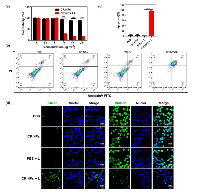
Given the superior photothermal properties of CR NPs, its PTT effectiveness in Colon26 and 4T1 was then studied (Fig. 3 and Determine S6). First, the photocytotoxicity of CR NPs was studied utilizing a normal CCK-8 cell viability assay. As proven in Fig. 3a, with out laser irradiation, the cell viability for Colon26 was negligibly influenced even the focus of CR NPs was as much as 40 µg mL− 1, revealing the superb biocompatibility of CR NPs for biomedical and scientific purposes. In distinction, important photothermal cytotoxicity was noticed after remedy with CR NPs + laser irradiation, and the cell viability decreased markedly with greater than 80% of cells dying at a CR NP focus of 20 µg mL− 1. Related outcomes had been additionally obtained in 4T1 cells, exhibiting that CR NPs + laser irradiation may considerably suppress the cell viability in a concentration-dependent method (Determine S6a). In the meantime, we observed that 4T1 cells seemed to be extra delicate to CR NPs-induced photothermal impact than Colon26 since decrease concentrations of CR NPs corresponding to 2.5 and 5 µg ml− 1 have urged outstanding inhibition to viability upon irradiation. To additional examine the photothermal impact of CR NPs, we then carried out reside (inexperienced fluorescence)/useless (pink fluorescence) staining experiments. As proven in Determine S6b, cells handled with PBS, CR NPs and PBS + laser irradiation exhibited widespread inexperienced fluorescence, indicating that there was no anticancer impact in these teams. Purple fluorescence was clearly noticed within the CR NPs + laser irradiation group, and nearly 70% of cells underwent loss of life after irradiation. This illustrates the superb PTT capability of CR NPs towards Colon26 cells, which is in step with CCK-8 findings. Moreover, cell apoptosis was studied by stream cytometry. As anticipated, the CR NPs + laser group confirmed a remarkably excessive cell apoptotic ratio (∼95%), whereas, within the management teams, no apparent apoptotic or necrotic cells had been noticed, which additional confirmed the excessive effectivity of the PTT impact of CR NPs (Fig. 3b and c). Collectively, CR NPs confirmed good biocompatibility and environment friendly PTT beneath laser irradiation, implying nice potential for in vivo biomedical purposes.
In vitro antitumor efficiency analysis of CR NPs. (a) Relative Colon26 cell viability in relation to varied CR NPs concentrations with or with out laser irradiation, decided utilizing a CCK-8 assay (n = 6). (b) Apoptosis evaluation on Colon26 cells utilizing stream cytometry after completely different therapies (CR NPs: 20 µg mL− 1). For (a) and (b), laser irradiation circumstances: 808 nm, 1.0 W cm− 2, 5 min. (c) The corresponding quantitative evaluation of (b). (d) Immunofluorescence staining of cell floor CALR and HMGB1 expression in numerous teams (n = 4). Scale bars = 50 μm
Research have indicated that photothermal ablation of tumors may stimulate and redistribute immune cells, thereby triggering sturdy antitumor immune responses [46]. An important side of this immune activation is the induction of ICD. ICD happens when tumor cells are subjected to exterior stimuli, mediating the physique’s antitumor immune response [47]. ICD is accompanied by the manufacturing of quite a lot of damage-associated molecular patterns (DAMPs), such because the publicity of surface-exposed calreticulin (CALR) and the discharge of high-mobility group field 1 (HMGB1) [48,49,50,51]. Right here, we investigated the expression of CALR on the cell floor and the discharge of HMGB1 in numerous teams. The PBS, CR NPs, and PBS + laser therapies confirmed little cell floor CALR publicity (inexperienced), whereas the CR NPs + laser remedy resulted in enhanced publicity of CALR on the cell floor owing to CR-mediated PTT beneath laser irradiation (Fig. 3d). As well as, the CR NPs + laser remedy group confirmed much less HMGB1 staining within the cell nuclei in contrast with the opposite three teams, indicating a outstanding launch of HMGB1. Collectively, these outcomes indicated that CR NPs can broadly induce tumor cell apoptosis and ICD after laser irradiation.
In vivo NIR-II fluorescence imaging and biodistribution evaluation of CR NPs
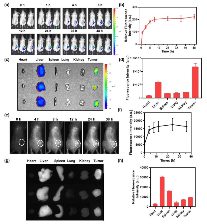
Contemplating the superb in vitro properties of the CR NPs, we then studied the imaging properties of the NPs in vivo. DIR-loaded NPs (CR/DIR NPs) had been constructed to watch the in vivo biodistribution and accumulation of CR NPs utilizing a Colon26 tumor-bearing mouse mannequin. CR/DIR NPs had been ready utilizing the identical technique as that used for the preparation of the CR NPs. When the tumor quantity reached a median measurement of ≈ 100 mm3, CR/DIR NPs (50 µg ml− 1, 100 µL, primarily based on DIR) had been injected intravenously into the mice. The biodistribution was examined utilizing IVIS at completely different time factors after injection. Because the whole-body pictures in Fig. 4a and b proven, fluorescent alerts had been primarily localized on tumor space and progressively enhanced over time, reaching a sign peak at 12 h after administration of CR/DIR NPs, which signifies wonderful tumor accumulation of the NPs. Notably, the fluorescent alerts of tumors handled with CR/DIR NPs remained clear and powerful even after 48 h, which supplied a very long time window for PTT software. Moreover, to guage the NPs distribution, ex vivo imaging of remoted primary organs and tumors was carried out 48 h after the injection. As proven in Fig. 4c and d, important fluorescence alerts had been nonetheless seen within the tumor and liver tissues 48 h after the injection of CR/DIR NPs, suggesting a robust passive tumor-targeting capability of CR/DIR NPs and liver clearance.
In vivo biodistribution evaluation and NIR-II FLI of CR NPs. (a) Imaging and (b) quantification evaluation of Colon26 tumor-bearing mice with CR/DIR NPs in numerous time factors (n = 3). (c and d) Ex vivo biodistribution and quantification of the dissected primary organs and tumors. (e) NIR-II fluorescence pictures and (f) the corresponding quantitative evaluation of CR NPs in 4T1 tumor-bearing mice at completely different occasions. 4T1 tumor-bearing mice had been intravenously injected with 100 µg of CR NPs (1.0 mg mL− 1) per mouse. Then, the real-time imaging was recorded and monitored at designated time intervals (0, 4, 8, 12, 24 and 36 h) post-injection (n = 3). NIR-II FLI circumstances: 808 nm, 1000 nm filter, 250 ms. White dashed circles point out tumor areas. (g) Ex vivo NIR-II FLI and (h) the corresponding quantification of main organs after 36 h administration of CR NPs
Inspired by these outcomes, we then evaluated the in vivo efficiency of CR NPs in NIR-II FLI utilizing a 4T1 tumor-bearing BALB/c mouse mannequin. First, the mice had been intravenously administered with CR NPs (100 µL, 1 mg mL− 1), and the NIR-II fluorescence pictures had been subsequently recorded at completely different occasions. Within the Fig. 4e and f, the tumor profile was simply distinguishable, indicating that CR NPs may successfully accumulate on the tumor web site. The NIR-II fluorescence intensities of the CR NPs at tumor areas progressively intensified inside 12 h and reached the plateau at 24 h after the injection, which was in accordance with the outcomes of NIR-I FLI (Fig. 4a and b). Due to this fact, the optimum time level for PTT is 12 h after the injection. Moreover, the fluorescent alerts had been maintained within the tumor area for over 36 h, indicating an extended therapeutic window for laser irradiation. Moreover, NIR-II ex vivo imaging of dissected tumors and very important organs was carried out 36 h after injection to additional consider the biodistribution of the CR NPs. As proven in Fig. 4g and h, the CR NPs had been primarily retained within the liver, spleen, and tumor, suggesting good tumor-targeting capability and potential metabolic organs. These imaging outcomes reveal that CR NPs possess favorable tumor-targeting capability, which is conducive to tumor remedy.
Notably, these promising knowledge reveal the potential for the applying of CR NPs in NIR-II imaging-guided surgical procedure. NIR-II FLI has emerged as a promising technique for exact image-guided tumor surgical procedure because of its deep tissue penetration and excessive spatial decision. The diminished background alerts, minimal tissue auto-fluorescence and low scattering within the NIR-II window allow clearer and extra exact imaging for tumor margins, which is essential for full tumor resection whereas minimizing injury to surrounding wholesome tissues. A number of research have proven the advantages of NIR-II FLI-guided most cancers surgical procedure, together with localizing cancers, evaluating surgical margins, guiding cytoreductive surgical procedure (CRS), tracing lymph nodes (LNs) and lymphatic vessels, and mapping particular anatomical buildings. These research have demonstrated the numerous prospects of NIR FLI guided surgical procedure in bettering surgical outcomes [52]. Moreover, the extended retention of CR NPs in tumor tissues offers a considerable therapeutic window, permitting surgeons adequate time to carry out image-guided surgical procedure. Total, CR NPs maintain important potential to enhance the outcomes of surgical interventions by means of very good tumor focusing on and imaging capabilities.
CR NPs-mediated in vivo photothermal remedy
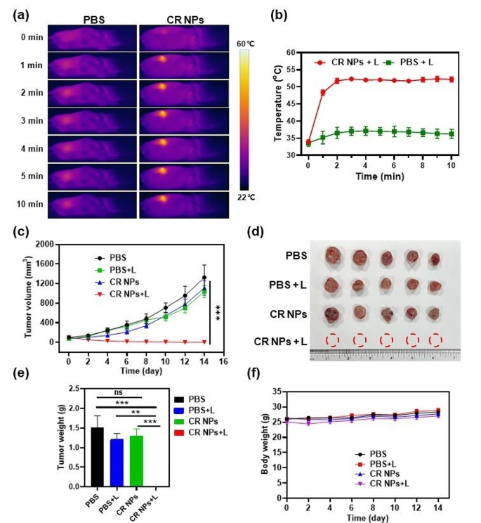
Primarily based on the environment friendly in vitro PTT impact and wonderful tumor accumulation of CR NPs, we then studied the PTT exercise of the CR NPs within the Colon26 tumor mannequin. When tumor measurement reached 100 mm3, the mice had been randomly divided into 4 teams (5 mice per group): (1) PBS, (2) PBS + laser, (3) CR NPs, and (4) CR NPs + laser. First, the mice had been intravenously injected with PBS and CR NPs; the tumors of mice in teams 2 and 4 had been then repeatedly irradiated for 10 min 12 h post-injection in accordance with the in vivo imaging knowledge. The actual-time photothermal pictures and temperatures of the mice within the laser-treated teams had been recorded with an IR thermal digital camera to verify the photothermal results of the CR NPs. Determine 5a and b illustrated that the tumor area’s temperature within the CR NPs + laser remedy group quickly elevated to 52 °C inside 2 min of irradiation and retained this temperature throughout the remaining interval of irradiation. The management group (PBS + laser) confirmed somewhat enhance in temperature (ΔT ≈ 4 °C) beneath the identical remedy circumstances, demonstrating that the photothermal impact induced by the laser alone was negligible and didn’t have a PTT impact in vivo.
In vivo CR NPs-mediated photothermal remedy. (a) Infrared thermal pictures and (b) corresponding temperature profiles on the tumor websites of Colon26 tumor-bearing mice handled with PBS or CR NPs beneath irradiation (808 nm, 1.0 W cm− 2) at completely different time factors (n = 5). (c) Tumor development profiles of 4 teams through the remedy course. Statistical significance was calculated through two-way ANOVA. (d-e) The tumor images (d) and (e) weights of dissected tumors of mice in every group on the 14th day. Statistical significance was calculated through one-way ANOVA. (f) Physique weight of mice among the many remedy teams. CR NPs injection and laser irradiation got solely as soon as
To additional consider the in vivo antitumor impact of CR NPs beneath laser irradiation, the tumor sizes and physique weights had been monitored each different day after remedy. Two days after PTT, black scars had been noticed within the tumor space from the CR NPs + laser group, whereas insignificant modifications had been discovered within the mice from the PBS + laser group. In Fig. 5c and Determine S7, the tumors of teams (1), (2) and (3) grew repeatedly with related excessive development charges, and the typical tumor quantity elevated to roughly 1500 mm3 inside 14 days. This means that CR NPs or laser irradiation alone don’t have any therapeutic impact. Notably, the NPs + laser remedy resulted in full tumor eradication. These outcomes point out a big in vivo photothermal therapeutic impact of CR NPs, which aligned with the in vitro findings. On the 14th day after remedy, the mice in all teams had been sacrificed and the tumors had been photographed and weighed (Fig. 5d and e). The tumor weight was in good settlement with the tendency of tumor development profiles, additional verifying the superb in vivo photothermal ablation impact of CR NPs. The physique weights of the mice had been related, with a slight enhance all through the remedy interval, demonstrating passable biocompatibility and restricted unwanted effects of CR NPs in vivo for PTT (Fig. 5f). Notably, a single-dose administration of CR NPs and as soon as irradiation had been carried out, which resulted in full tumor elimination, additional demonstrating the superior PTT efficacy of CR NPs for in vivo remedy.
To additional consider the biosafety of CR NPs, histological staining of main organs and blood biochemical evaluation of the handled mice had been carried out on the finish of remedy. As proven in Determine S8, the histological evaluation of the most important organs of all mice confirmed no important tissue injury or irritation after remedy, additional confirming the superb in vivo biosafety of the CR NPs. The physiological security of the CR NPs was estimated utilizing routine blood and biochemistry evaluation (Determine S9). Numerous blood parameters confirmed no important variations among the many 4 teams after remedy, indicating that the CR NPs didn’t present systemic toxicity or dangerous impacts on livers and kidneys of the mice. Taken collectively, CR NPs are a promising candidate for exact NIR-II FLI-guided PTT in vivo, with wonderful theranostic functionality and negligible unwanted effects.
Antitumor immune reminiscence evaluation
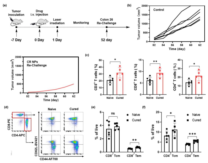
Given the whole tumor eradication within the Colon26 tumor mannequin after remedy with CR NPs + laser irradiation, we questioned whether or not antitumor reminiscence could possibly be induced by CR NPs. After remedy, tumor-free mice in one other batch of mice had been re-challenged with 2.0 × 10⁶ Colon26 tumor cells on day 52 after the preliminary remedy (Fig. 6a). Age-matched mice injected with Colon26 cells (2 × 10⁶) cells had been used as controls. The volumes of the tumors had been measured each alternate day. Consequently, the 4/5 of mice didn’t kind tumors within the re-challenged mice receiving the remedy of CR NPs + laser irradiation, whereas all six mice within the management group developed quickly rising tumors (Fig. 6b), indicating that antitumor immune reminiscence is developed after publicity to CR NPs. To elucidate the basic mechanism, stream cytometry was used to quantify the memory-associated T cells within the spleen. The outcomes of Fig. 6c and Determine S10 indicated that CD3+, CD3+CD8+, and CD3+CD4+ T lymphocytes within the spleens of rechallenge-resistant mice had been notably elevated when put next with the management group. The stream cytometric evaluation additional revealed that effector reminiscence T cells (Tem, CD62L+CD44−) had been noticeably elevated each in CD4+ and CD8+ T cells, and central reminiscence T cells (Tcm, CD62L+CD44+) in CD4+ T cells had been additionally considerably elevated (Fig. 6d–f). These outcomes clearly point out that the CR NPs + laser remedy can successfully inhibit tumor development and promote the formation of long-term antitumor reminiscence, thereby probably stopping tumor metastasis.
Antitumor immune reminiscence evaluation. (a) Mice cured by CR NPs + laser remedy had been re-challenged by Colon26 cells (2 × 10⁶ cells) on day 52 after the preliminary remedy. (b) Tumor quantity of every re-challenged mouse. (c) Quantification of CD3+, CD3+CD8+, CD3+CD4+ T lymphocytes within the spleen. (d-f) Move cytometric (FCM) evaluation and quantitative knowledge of central reminiscence T (Tcm, CD62L+CD44+) cells and effector reminiscence T (Tem, CD62L−CD44+) cells subset from CD8+ and CD4+ T cells within the spleen. Knowledge are proven because the imply ± SD, n = 5–6. Statistical significance was calculated through a two-tailed Pupil’s t-test
Anti-primary and metastatic tumor efficacy of CR NPs in 4T1 breast most cancers mannequin
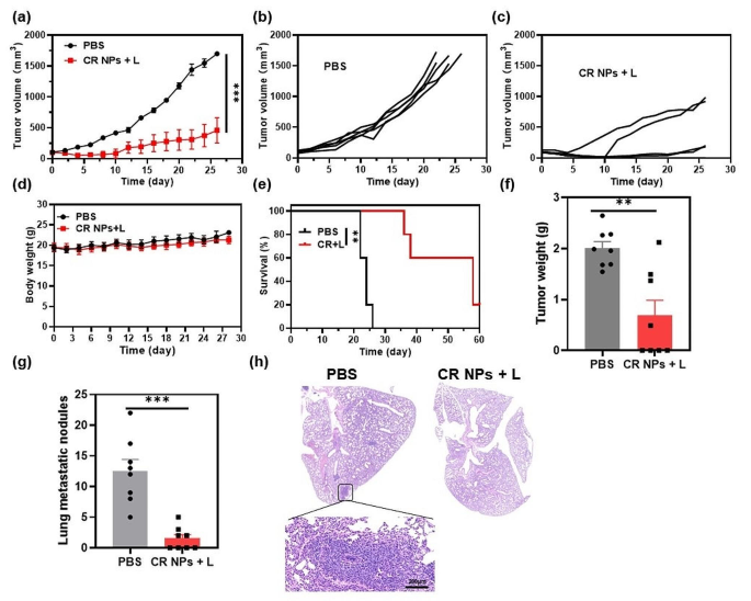
Given the induction of long-term antitumor immune reminiscence in Colon26 tumor-bearing mice, we additional investigated whether or not CR NPs + laser irradiation may exert concurrently anti-primary and anti-metastatic tumor results utilizing an orthotopic 4T1 murine mammary carcinoma mannequin which may induce lung metastasis throughout main tumor improvement. We first assessed the anti-primary tumor efficiency on this refractory tumor mannequin. When the tumor volumes reached 100 mm3, the mice had been handled with PBS or CR NPs + laser irradiation 12 h after injection. The tumor volumes and physique weights had been recorded each different day through the remedy interval. The outcomes confirmed that CR NPs + laser irradiation successfully inhibited orthotopic 4T1 tumor development, though no tumor eradication was noticed owing to its resistance to remedy (Fig. 7a–c), demonstrating the vast applicability of CR NPs-mediated PTT in numerous tumor varieties. Equally, CR + laser remedy didn’t lead to any important alterations in animal physique weight in comparison with PBS remedy (Fig. 7d). The survival time of the mice within the CR NPs + laser remedy group was additionally drastically extended (Fig. 7e), whereas tumors in mice handled with PBS alone grew quickly, with a median lifespan of solely 20–26 days. These outcomes counsel that CR NPs + laser irradiation is extremely environment friendly and secure for the in vivo photothermal ablation of extremely metastatic tumors.
As most cancers metastasis is the principle reason behind tumor-associated loss of life, we subsequently investigated whether or not CR NPs + laser irradiation may scale back lung metastasis of 4T1 tumors after remedy in one other batch of tumor-bearing mice. Tumor development of mice within the CRs + laser group was drastically inhibited, which was in keeping with the outcomes of the primary batch of mice (Determine S11). The tumors and lungs of the mice had been collected on day 30 after remedy to guage the antimetastatic results. As displayed in Fig. 7f and g, the first tumor weight was considerably decrease in CR NPs + laser-treated mice than in management mice, and the variety of pulmonary metastatic tumor nodules on the lung floor was additionally remarkably diminished in handled mice when put next with the management group. The lungs had been sectioned for histological analysis, and nearly no metastatic lesions had been noticed within the CR NPs + laser-treated group (Fig. 7h), whereas important metastatic foci had been found within the management group’s lungs, indicating that CR NPs-based PTT successfully inhibits the lung metastasis of tumor cells. Due to this fact, the CR NPs-based PTT can induce a potent systemic immune response and stop tumor metastasis.
Anti-primary and metastatic tumor efficacy on a 4T1 breast most cancers mannequin through the use of CR NPs. (a) Tumor quantity development curves from the 2 handled teams (n = 5). Statistical significance was calculated through two-way ANOVA. Corresponding tumor development curves in (b) PBS and (c) CR NPs + laser teams. (d) Physique weights of the handled mice. (e) Survival curves (n = 5). Statistical significance was calculated through Log-rank (Mantel-Cox) take a look at. (f) Tumor weights and (g) the variety of metastatic nodules on lung floor on the endpoint. Statistical significance was calculated through unpaired Pupil’s-test. (h) Typical hematoxylin and eosin (H&E) staining of lungs from the 2 teams


