Characterization of 78c@Lipo-FA
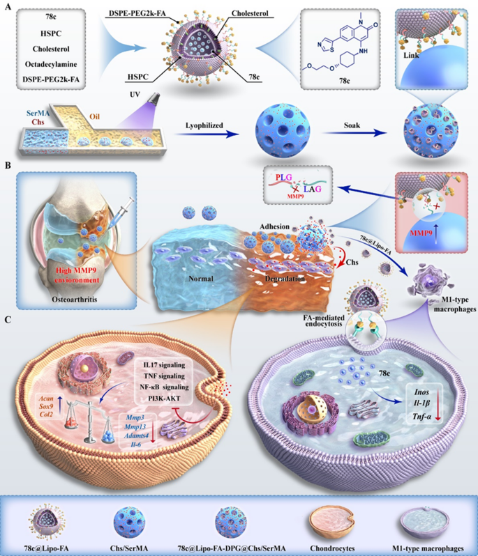
As proven in Scheme 1, 78c@Lipo-FA was ready by including DSPE-PEG2k-FA and 78c proportionally utilizing a thin-film dispersion methodology. Chs/SerMA hydrogel microspheres had been ready by microfluidics, and the MMP9-responsive peptide DPG was covalently connected to the Chs/SerMA hydrogel microspheres by maleimide and sulfhydryl reactions. Lastly, 78c@Lipo-FA, because the secondary construction of microspheres, was ready to hyperlink with DPG@Chs/SerMA hydrogel microspheres to kind LFDCS (Scheme 1A).
Bionic bearing-inspired lubricating microspheres characterised by immunomodulatory for osteoarthritis therapy. (A) Synthesis of M1-targeted liposomes and extremely adhesive lubricating hydrogel microspheres. (B) Utility of composite hydrogel microspheres in OA. (C) Mechanism of composite hydrogel microspheres to ameliorate chondrocyte dysfunction by downregulation of inflammatory and PI3K/AKT signaling pathways and inhibit M1-mediated irritation. Ultraviolet: UV
78c, as a CD38 inhibitor, not solely inhibits macrophage operate by suppressing CD38 signaling in M1 macrophages, but in addition reveals nontoxicity to M2 macrophages, indicating a superb therapeutic prospect for concentrating on irritation in OA [30]. In line with earlier findings, we discovered that the M1 macrophage inhabitants was considerably decrease in LPS + 78c group ( 51.8%) as in comparison with the LPS group (77.4%) (Determine S1A) [30]. This conclusion was supported by quantitative real-time PCR (qRT-PCR) outcomes for the gene expression of Tnf-α, Il-1β, and Inos (Determine S1B). Nevertheless, 78c is a fat-soluble compound with low solubility in aqueous answer and is shortly cleared by the synovial fluid, severely lowering the intra-articular drug supply effectivity [38]. As a drug supply system, liposomes exhibit the power to reinforce drug loading by the lipophilic properties of phospholipids, thereby selling the efficient uptake of medicine by cells; consequently, they’ve supplied wonderful drug supply within the medical setting [39].
The folic acid receptor (FAR) household encompasses 4 members, particularly FARα, β, γ, and δ [40]. As a receptor broadly expressed upon M1 macrophages, FAR is basically absent in M0, M2 macrophages, and regular cells [41]. Moreover, giant numbers of FAR-positive macrophages are current in OA [42]. Subsequently, we used FA as a ligand to switch liposomes (Lipo-FA) to successfully goal M1 macrophages, which didn’t have an effect on the remaining cells.
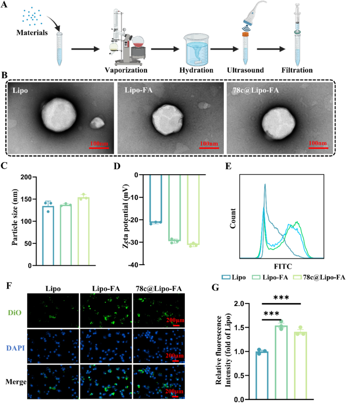
Within the earlier research, liposomes had been preparation by thin-film dispersion methodology. As illustrated in Fig. 1A, liposome movies had been ready utilizing a rotary evaporator after mixing the supplies in a centrifuge tube. 78c@Lipo-FA was ready by stirring, ultrasonicating, and extruding the liposome suspension. The TEM outcomes revealed that the liposomes had a uniform spherical look (Fig. 1B). The diameters of Lipo, Lipo-FA, and 78c@Lipo-FA had been 134.53 ± 11.03 nm, 137.23 ± 2.67 nm, and 154 ± 5.97 nm, respectively (Fig. 1C), and exhibited nicely dispersion (Determine S1C). The zeta potentials of Lipo, Lipo-FA, and 78c@Lipo-FA had been -21.3 ± 0.49 mV, -46.2 ± 0.95 mV, and -31.2 ± 0.79 mV, respectively (Fig. 1D, Determine S1D). Apparently, we discovered that after FA modification, the zeta potential of the liposomes elevated roughly 2-fold, which can be as a result of anchoring of the negatively charged useful teams current within the DSPE-PEG2k-FA band to the liposomes [43]. Moreover, the Lipo-FA encapsulation of 78c was roughly 74% (Determine S2).
To check the concentrating on of Lipo-FA, movement cytometry assay was employed to evaluate the uptake of liposomes by BMMs. The outcomes revealed that, in comparison with Lipo alone, Lipo-FA and 78c@Lipo-FA had been taken extra by M1 macrophages and the loading of 78c had no vital impact on the concentrating on of liposomes (Fig. 1E), suggesting that the modification of FA enhance the uptake of liposomes. Noticed by a fluorescence microscope, as depicted in Fig. 1F and G, M1 macrophages displayed a better uptake of Lipo-FA and 78c@Lipo-FA than the Lipo group, and the fluorescence intensities of Lipo-FA and 78c@Lipo-FA had been 1.5 occasions and 1.4 occasions increased than these of the Lipo group, additional confirming the concentrating on of Lipo-FA. In sum, we designed FA-modified liposomes able to monitoring and delivering medicine to M1 macrophages, thus enhancing the liposome supply effectivity [44].
Characterization of 78c@Lipo-FA. (A) Scheme diagram of the preparation means of Lipo-FA; supplies together with 78c, HPSC, ldl cholesterol, octadecylamine, and DSPE-PEG2k-FA. (B) TEM of liposomes. (C) Particle measurement of liposomes (n = 3). (D) Zeta potential of liposomes (n = 3). (E) Consultant panels of the uptaking-liposomes within the BMMs assayed by movement cytometry. (F) Consultant photos of the uptaking-liposomes within the BMMs noticed by the fluorescence microscope (After BMMs had been incubated for 4 h). (G) Relative fluorescence depth of the uptake of liposomes by BMMs (n = 3). ***p < 0.001
Characterization and lubrication properties of LFDCS
SerMA hydrogel reveals completely different traits at completely different concentrations [45, 46]. To find out the acceptable focus of SerMA to arrange the microspheres, we examined the G’ and G’’ concentrations of the SerMA options in the course of the curing course of with a rheometer. As proven in Determine S3, we discovered that completely different concentrations of options fused nicely after photo-crosslinking, and 25% w/v SerMA had increased viscosity than the opposite teams. Subsequently, 25% w/v SerMA was chosen for subsequent experiments.
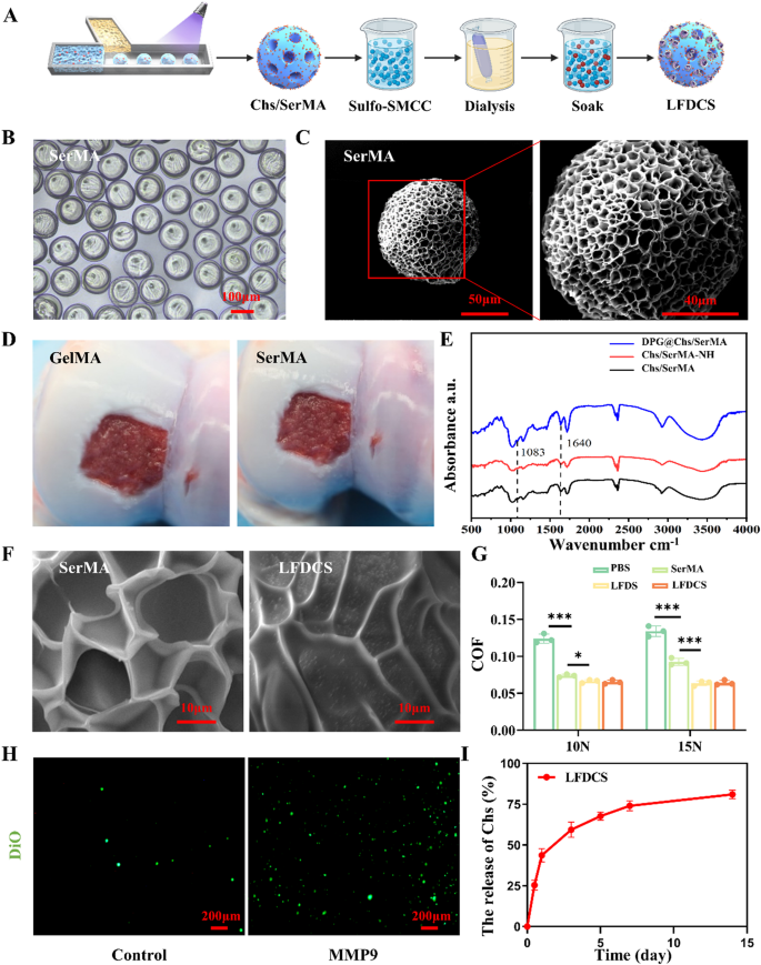
Chs/SerMA hydrogel microspheres had been fabricated with a microfluidic machine, and the amino teams on the Chs/SerMA hydrogel microspheres had been activated by Suflo-SMCC. Subsequently, DPG was covalently connected to the activated Chs/SerMA hydrogel microspheres. After purification by dialysis, 78c@Lipo-FA was certain to DPG@Chs/SerMA hydrogel microspheres by way of DSPE segments embedded within the hydrophobic area of liposomes (Fig. 2A). SerMA was chosen because the service due to the traits: firstly, sericin accommodates numerous reactive teams delicate to chemical modifications; secondly, sericin possesses excessive adhesion properties [10, 12, 47]. After demonstrating the wonderful bodily adhesion properties of 25% w/v SerMA, the microstructure of the microspheres was studied. Fig. 2B confirmed that the SerMA hydrogel microspheres displayed wonderful spherical form and homogeneity. Scanning electron microscopy (SEM) revealed that SerMA hydrogel microspheres had a porous construction with a diameter of about 100 μm (Fig. 2C).
Presently, microsphere preparation supplies reminiscent of Chs methacrylate, HA methacrylate, and GelMA, that are generally employed within the therapy of OA, require chemical modifications to reinforce their adhesion properties for cartilage tissue engineering due to their inherently weak adhesion potential [3, 4]. To analyze the affect of SerMA hydrogel microspheres on cartilage adhesion, GelMA hydrogel microspheres had been utilized because the management group [48]. The outcomes indicated that the microspheres of 25% w/v SerMA hydrogel microspheres exhibited superior adhesion to cartilage and defect websites in comparison with the microspheres of seven.5% w/v GelMA hydrogel microspheres (Fig. 2D, Supplementary Video 1). Thus, SerMA-based microspheres might successfully adhere to the cartilage floor with out further modification.
FTIR spectroscopy and SEM had been carried out to verify the binding of 78c@Lipo-FA to SerMA hydrogel microspheres. The outcomes of FTIR spectroscopy indicated the presence of amide and ether bands at 1,680 cm− 1 and 1,083 cm− 1 in DPG@Chs/SerMA in comparison with Chs/SerMA and activated Chs/SerMA. (Chs/SerMA-NH) (Fig. 2E). We additionally noticed the floor morphologies of SerMA, LFDS, and LFDCS by SEM. As proven in Fig. 2F and Fig. S4, some dispersed liposome microreservoirs had been noticed on the surfaces of LFDS and LFDCS, whereas they had been absent in Chs/SerMA or SerMA hydrogel microspheres, suggesting that Lipo-FA are connected to the microspheres.
To discover the lubrication properties of the microspheres, SerMA, LFDS, and LFDCS had been positioned below 10 N and 15 N hundreds within the common materials testers, respectively. The outcomes confirmed that the coefficients of friction (COF) of SerMA, LFDS, and LFDCS had been diminished in contrast with PBS, with the COF values of LFDS and LFDCS reducing by roughly 49% and 48%, respectively (Fig. 2G, Fig. S5). Owing to the excessive viscosity and rolling traits, SerMA hydrogel microspheres can improve lubrication by appearing as ball bearings [49]. As well as, the leads to Fig. 2G revealed that the lubricating properties of LFDS and LFDCS had been superior to these of SerMA, indicating that the hydrated synovial membrane supplied by liposomes might additionally scale back the harm to the cartilage by mechanical friction and additional synergistically scale back the COF of the interface [36].
Systemic or native injection of focused liposomes typically leads to unsatisfactory therapeutic results due to their brief half-lives and restricted distances of motion [50]. Subsequently, it’s crucial to ascertain a liposome preservation reservoir that may protect liposomes and is programmed to manage their launch [51]. Irregular inflammatory states and cartilage friction enhance the secretion of IL-6, prostaglandin E2, and MMP3, MMP9, and MMP13 inside the joints [52]. On this research, LFDCS had been designed as a conserved financial institution of 78c@Lipo-FA. Upon the presence of MMP9, reminiscent of within the microenvironment of the OA and trauma, LFDCS might launch 78c@Lipo-FA with DPG cleaved by MMP9 for therapeutic functions.
As proven in Determine S6, the attribute peaks of DSPE, PEG2k, and GPLGLAGQC had been noticed within the nuclear magnetic resonance consequence, indicating profitable synthesis of DPG. Subsequent, DiO dyed-liposomes had been noticed to check the launched 78c@Lipo-FA from LFDCS. In vitro, as anticipated, extra liposomes with inexperienced fluorescence had been discovered of LFDCS within the MMP9 atmosphere than in management group with out MMP9 (Fig. 2H).
Chs is a crucial structural element of the cartilage extracellular matrix (ECM) that promotes chondrocyte anabolism and inhibits chondrocyte catabolism to facilitate cartilage restore [53]. On this research, we ready SerMA hydrogel microspheres combined with Chs. First, the focus of Chs within the LFDCS launch answer was decided with HPLC. The outcomes demonstrated that Chs had an preliminary burst launch inside 24 h, adopted by a gradual plateau, reaching a plateau at 7 d, which displays roughly 80% encapsulation of Chs (Fig. 2I, Fig. S2). The in vitro degradation profile revealed that LFDCS confirmed continued biodegradability (Determine S7).
In abstract, LFDCS adheres to cartilage and offers lubrication to joints, whereas additionally responds to MMP to launch liposomes.
Characterization of LFDCS. (A) Scheme diagram of the preparation of LFDCS. (B) The morphology of SerMA hydrogel microspheres noticed by the sunshine microscope. (C) The consultant SEM photos of SerMA hydrogel microspheres. (D) The gross statement of GelMA and SerMA hydrogel microspheres adhension to porcine cartilage. (E) FTIR of DPG@Chs/SerMA, Chs/SerMA-NH, and Chs/SerMA. (F) The consultant SEM picture of SerMA and LFDCS. (G) COF histograms of PBS, SerMA, LFDS, and LFDCS (n = 3). (H) The responsive launch of liposomes in LFDCS noticed by the fluorescence microscope. (I) Launch of Chs in LFDCS (n = 3). *p < 0.05, *** p < 0.001
Biocompatibility of LFDCS
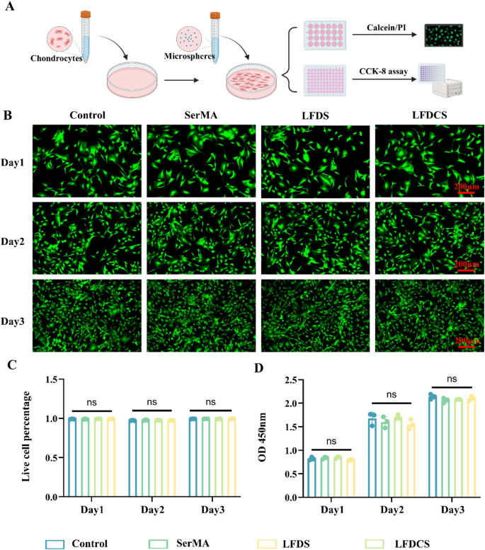
To evaluate the biocompatibility of the microspheres, rat chondrocytes had been efficiently remoted evidenced by toluidine blue staining (Determine S8) and cultured within the leachate from every group (SerMA, LFDS, and LFDCS, respectively). Calcein/PI staining and CCK-8 had been utilized to find out the toxicity of the microspheres to chondrocytes at day 1, 2, and three, respectively (Fig. 3A). Calcein/PI staining and quantification outcomes demonstrated that there was no vital distinction between the teams on the similar timepoints (Fig. 3B, C). The CCK-8 assay displayed that the optical density values of the biomaterial group had been similar to these of the management group (Fig. 3D), additional confirming that the biocompatibility of LFDCS was good and appropriate for making use of in vivo.
LFDCS inhibited macrophage polarization towards M1 phenotype
Synovial macrophages are pivotal in producing pro-inflammatory components (IL-1β and TNF-α) following joint damage [54]. Within the synovium of the knee joints of sufferers with OA, there’s a vital activation of M1-mediated irritation in contrast with wholesome people [55]. Regardless of the secretion of IL-4 and IL-10 by M2 macrophages, these components are inadequate to counteract inflammatory infiltration, cartilage degradation, and bone regrowth attributable to M1 macrophages [22, 56]. Subsequently, modulation of intra-articular M1-mediated irritation is a vital technique for managing OA. As talked about beforehand, the inhibition of macrophage CD38 expression successfully inhibited macrophage polarization towards M1 phenotype (Determine S1A).
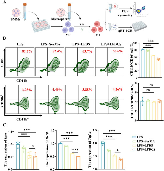
To look at the affect of LFDCS on macrophage polarization, 100 ng/mL LPS was utilized to induce polarization towards M1 phenotype (Fig. 4A). As proven within the Fig. 4B, the proportion of CD11b+CD86+ (M1 phenotype) BMMs within the LFDS and LFDCS teams decreased by roughly 17.4% and 27.5%, respectively, compared to the LPS group, whereas the proportion of CD11b+CD206+ (M2 phenotype) BMMs was under 6% in each teams.
The expression of Inos within the LFDS and LFDCS teams decreased by 0.43 occasions and 0.46 occasions, the expression of Il-1β by 0.39 occasions and 0.49 occasions, and the expression of Tnf-α by 0.48 occasions and 0.62 occasions, respectively, compared to the LPS group (Fig. 4C). Moreover, the proportion of M1 macrophages and the expression of associated genes within the LFDCS group had been increased than that within the LFDS group, which can be attributable to Chs inhibiting irritation in M1 macrophages by eliminating ROS [53].
LFDCS inhibited macrophage polarization towards M1 phenotype. (A) Scheme diagram of microspheres intervention on macrophage polarization. (B) The portion of CD11b+CD86+ (M1 phenotype) and CD11b+CD206+ (M2 phenotype) detected by movement cytometry at 24 h (n = 3). (C) The gene expression of M1 polarization-related genes (Inos, Il-1β and Tnf-α) assayed by qRT-PCR at 24 h (n = 3). *p < 0.05, ***p < 0.001, and ns, no vital distinction
LFDCS ameliorated IL-1β-induced chondrocyte dysfunction
Irritation-induced hypertrophic deformation of chondrocytes is a vital indicator of OA [57]. Subsequently, 10 ng/mL IL-1β was used to elicit inflammation-induced chondrocyte deformation in vitro [58]. ACAN, Col2, and SOX9 are essential components in chondrocyte regeneration and ECM synthesis, whereas MMP3, MMP13, and ADAMTS4 regulate chondrocyte catabolism [59].
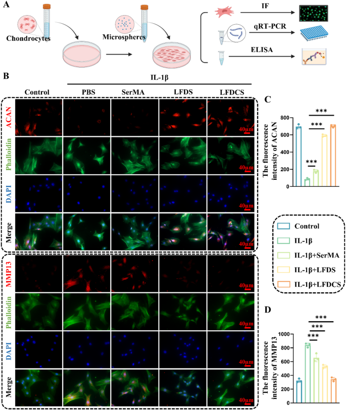
Firstly, rat chondrocytes had been cultured for twenty-four h within the leachate from every group, and useful indicators within the chondrocytes had been detected by IF staining and qRT-PCR (Fig. 5A). The Fig. 5B demonstrated a gradual enhance in ACAN expression and a gradual lower in MMP13 expression within the chondrocytes within the SerMA, LFDS and LFDCS group, in contrast with that of the IL-1β group. Quantitative outcomes displayed the expression of ACAN up-regulated 1.06-, 5.84-, and seven.1-fold, whereas the expression of MMP13 down-regulated by 28%, 59%, and 144%, within the SerMA, LFDS, and LFDCS teams, in contrast with that within the IL-1β group, respectively (Fig. 5C and D).
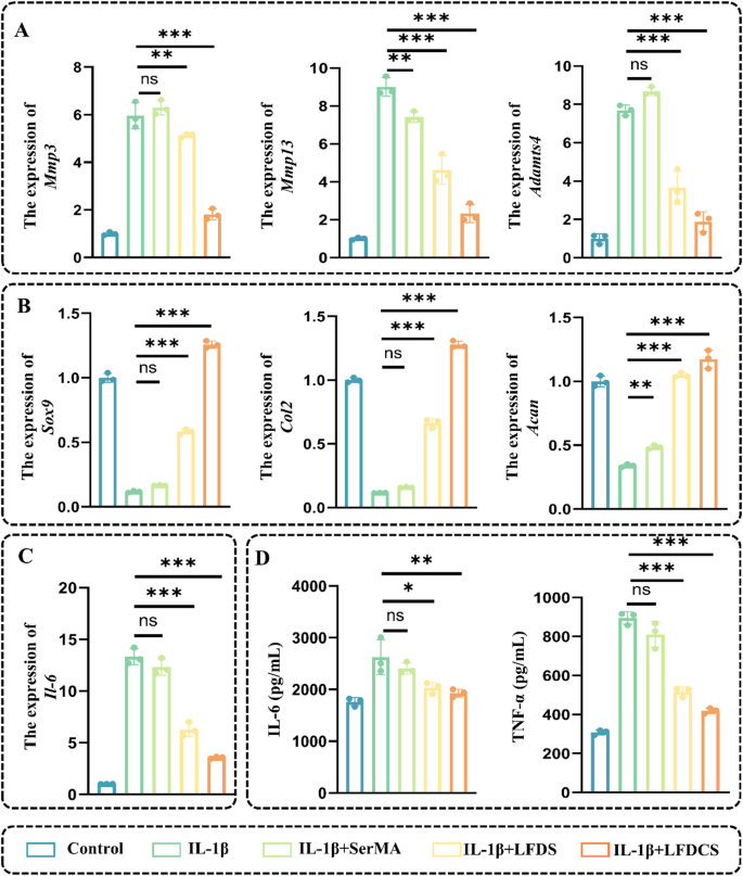
As well as, chondrocyte synthesis and catabolism had been additional assessed by qRT-PCR. In distinction to the outcomes from the IL-1β group, there was a considerably declined gene expression of chondrocyte catabolism-related markers, together with Mmp3 (decreased by 14% and 70%, respectively), Mmp13 (decreased by 48% and 74%,, respectively), and Adamts4 (decreased by 52% and 76%, respectively) and a considerably enhanced gene expression of chondrocyte synthesis-related markers together with Sox9 (elevated 4.27- and 10.36-fold, respectively), Col2 (elevated 5- and 10.54-fold, respectively), and Acan (elevated 2.05- and a couple of.44-fold, respectively) within the LFDS and LFDCS teams. Chs is a vital structural element in cartilage and performs an vital position in sustaining the cartilage-forming phenotype [53]. The above outcomes indicated that the sluggish launch of Chs in LFDCS group considerably alleviated chondrocyte dysfunction in comparison with LFDS group.
LFDCS up-regulated ACAN and down-regulated MMP13 expression in chondrocytes. (A) Scheme diagram of LFDCS intervention in IL-1β-induced chondrocytes. (B) The expression of ACAN and MMP13 within the chondrocytes detected by IF. (C, D) Quantification of ACAN and MMP13 expression within the chondrocytes (n = 3). *** p < 0.001
In the meantime, the inflammatory response was additionally evaluated. In comparison with the IL-1β group, a lower within the gene expression of Il-6 (decreased by 53% and 73% within the LFDS and LFDCS teams, respectively) was noticed by qRT-PCR (Fig. 6A–C). Within the preliminary phases of OA, chondrocytes reactively secrete TNF-α and IL-6 to exacerbate OA development [60]. Moreover, ELISA outcomes revealed that the secretion of IL-6 and TNF-α of chondrocyte had been dropped within the LFDS and LFDCS teams, in comparison with that within the IL-1β teams (Fig. 6D). Apparently, the LFDS group had the power to advertise cartilage operate in comparison with the SerMA group (Fig. 5B). This can be on account of the truth that the 78c launched from the leachate of the LFDS group straight acted on the chondrocytes to ameliorate chondrocyte operate [29, 30].
LFDCS ameliorated IL-1β-induced chondrocyte dysfunction. (A) The expression of chondrocyte catabolism-related genes (Mmp3, Mmp13 and Adamts4) detected by qRT-PCR. (B) The expression of chondrocyte anabolism-related genes (Sox9, Col2 and Acan) detected by qRT-PCR. (C) The expression of chondrocyte inflammatory gene Il-6 detected by qRT-PCR. (D) IL-6 and TNF-α focus detected by ELISA. * p < 0.05, ** p < 0.01, *** p < 0.001, and ns, no vital distinction
LFDCS attenuated fluid-phase signaling and diminished osteoid formation within the joints of OA rats
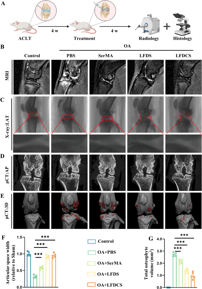
In vitro, we verified that LFDCS promoted cartilage adhesion, attenuated M1 macrophage irritation, and ameliorated chondrocyte dysfunction; due to this fact, we utilized LFDCS in rats with anterior cruciate ligament transection (ACLT) [61]. One month after surgical procedure, rats had been randomly divided and injected with PBS and the microspheres (SerMA, LFDS, and LFDCS, respectively) within the articular house and recorded because the PBS, SerMA, LFDS, and LFDCS teams, whereas the rats with sham surgical procedure had been set because the management group. After 4 weeks of therapy, the rats had been scanned with MRI and µCT, and their joints had been collected for histological staining (Fig. 7A). The in vivo compatibility take a look at revealed that the injection of microspheres had no vital impact on the inner organs in every group (Fig. S9A).
To discover the retention of LFDCS within the joint cavity, Cy5.5 was loaded onto LFDCS, and the change of fluorescence sign within the joint cavity after surgical procedure was noticed by an in vivo imaging system. One month after ACLT, with intra-articular injection of Cy5.5-labled LFDCS, the fluorescence sign within the joint cavity was maintained at a comparatively secure degree at first, and steadily decreased with the gradual degradation of LFDCS. Until to the 14th day, a fluorescence sign was nonetheless seen within the joint cavity, indicating that LFDCS have a superb slow-release functionality within the joint cavity (Fig. S9B).
In keeping with the MRI outcomes, the fluid-phase alerts within the joint cavities of the rats within the PBS, SerMA, LFDS, and LFDCS teams steadily decreased in contrast with that of the management group (Fig. 7B). And the fluid-phase alerts of LFDCS had been just like that of the management group, which indicated that the administration of LFDCS tremendously diminished joint edema. Narrowing of the joint house on radiographs often signifies OA with progressive cartilage harm [62]. Lateral knee X-rays revealed that the articular house width within the PBS group decreased 2.13-fold in contrast with the management group, whereas the articular house width within the SerMA group elevated 0.81-fold, the LFDS group elevated 1.88-fold, and that within the LFDCS group elevated 1.97-fold in contrast with the PBS group (Fig. 7C and F). In keeping with the anteroposterior view of the knee joint, medial subchondral osteosclerosis was considerably extra extreme within the PBS group than within the management group. Nevertheless, it was alleviated by being handled with SerMA, LFDS, and LFDCS, in contrast with that of the PBS group (Fig. 7D).
Intraarticular osteophyte formation is one other pathogenic function of OA [63]. The outcomes of µCT and 3D reconstruction indicated that the diploma of intra-articular osteophyte formation was higher within the PBS group than within the management group. Nevertheless, the diploma of osteophyte formation was alleviated within the SerMA, LFDS and LFDCS teams (Fig. 7E, G). In keeping with the outcomes proven in Fig. 7B-E, the advance of OA by LFDCS was considerably higher than that within the LFDS and SerMA teams, which was in line with the leads to the in vitro experiments. It recommended that LFDCS not solely forestall additional joint harm by lowering joint friction, but in addition promotes the restore of already broken cartilage by slowly releasing Chs.
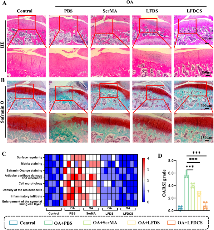
LFDCS diminished fluid-phase signaling and osteoid formation within the joints of OA rats. (A) Scheme diagram of LFDCS therapy. (B) Edema within the joint cavity detected by MRI scanning. (C) Articular surfaces detected by X-ray scanning, LAT: lateral. (D) Articular surfaces detected by µCT scanning, AP: anteroposterior. (E) 3D reconstruction of the knee joint by µCT scanning. (F) X-ray quantification of the medial cartilage hole width in rats (n = 5). (G) Quantification of whole osteophyte within the rat knee joint (n = 5). *** p < 0.001
LFDCS diminished cartilage harm in OA rats
For histological evaluation of the cartilage, the management group had clean cartilage surfaces and uniform ECM. The PBS group had extreme cartilage defects and irregular mobile association. In distinction, these impairments had been mitigated within the SerMA, LFDS, and LFDCS teams in contrast with the PBS group (Fig. 8A). Notably, though the diploma of cartilage put on was related within the SerMA and PBS teams, the SerMA group had a extra full cartilage construction and extra neatly organized cells than the PBS group did. Thus, SerMA hydrogel microspheres injection is useful for delaying OA development.
Safranin O staining was carried out on rat cartilage sections to distinguish cartilage and bone. Extra extreme cartilage defects and fibrous hyperplasia across the articular surfaces had been noticed in PBS group than within the management group. Akin to the outcomes of radiology and H&E staining, cartilage put on considerably improved within the SerMA, LFDS, and LFDCS group in contrast with the PBS group (Fig. 8B). In comparison with the PBS, SerMA and LFDS teams, the cartilage across the articular surfaces within the LFDCS group was extra evenly aligned, with much less cartilage put on and fewer fibroplasia (Fig. 8B). The microstructure of the repaired cartilage was graded by the use of a histological rating to quantify the diploma of cartilage therapeutic. The outcomes demonstrated a gradual enhance in cartilage restore scores with therapy within the SerMA, LFDS, and LFDCS teams in comparison with the PBS group (Fig. 8C). The OARSI scores for the PBS, LFDS, and LFDCS teams had been roughly 5.73, 2.73, and 0.8, respectively, confirming that LFDCS can considerably delay OA development (Fig. 8D).
LFDCS ameliorated cartilage dysfunction and attenuated synovial irritation in OA rats
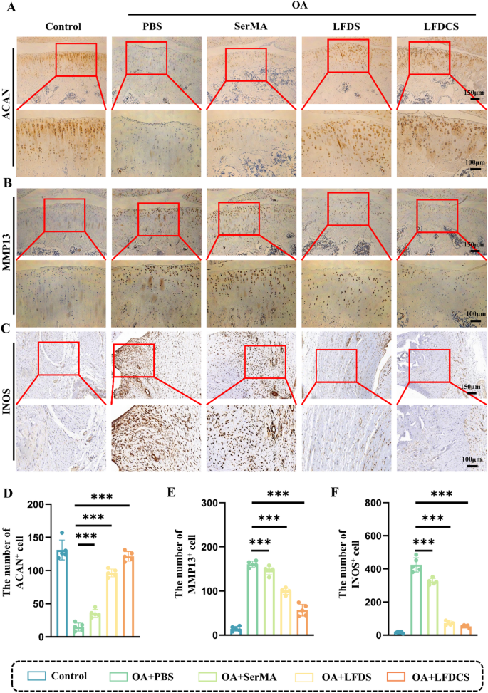
We additional analyzed the expression of ACAN and MMP13 on the cartilage floor by IHC evaluation. As proven in Fig. 9A, B, D, E, there was a 1.39-fold enhance within the expression of ACAN within the SerMA group, a 5.5-fold within the LFDS group, and a 7.21-fold within the LFDCS group, whereas the expression of MMP13 was diminished by 8% within the SerMA group, 38% within the LFDS group, and 65% within the LFDCS group in comparison with the PBS group. Intraarticular injection of LFDCS considerably promoted the expression of ACAN and suppressed the expression of MMP13 on the joint floor (Fig. 9A, B). That is in line with our hypothesis within the in vitro a part of the experiment. 78c within the LFDS group principally acted on M1 macrophages, and the sluggish launch of Chs in LFDCS additional ameliorated chondrocyte dysfunction when subjected to extrusion. Apparently, the upper ACAN and the decrease MMP13 within the SerMA group than within the PBS group could also be defined by the truth that SerMA hydrogel microspheres act as a bionic bearing to scale back cartilage put on and chondrocyte harm, thereby bettering chondrocyte metabolism. In abstract, LFDCS can be utilized as each a biolubricant and a drug supply automobile to attenuate cartilage put on and tear, reduce osteophyte, and promote ECM manufacturing, thereby assuaging OA development. As well as, to additional confirm the movement cytometry outcomes of Fig. 4, we analyzed the expression of INOS on the synovium of rat joints by IHC. As proven in Fig. 9C, F, the expression of INOS was decreased within the LFDS and LFDCS teams in contrast with the PBS group, which indicated that the intervention of 78c considerably suppressed macrophage irritation. In sum, it’s illustrated that below the high-MMP9 atmosphere of OA, many of the 78c@Lipo-FA launched from the LFDCS exerted a job in concentrating on M1 macrophages, whereas Chs situated inside the LFDCS nicely exerted a job in ameliorating chondrocyte dysfunction.
The pathology of OA is often associated to insufficient vascular perfusion, irritation, and decreased lubrication [22, 64]. The interplay between recurrent inflammatory responses and cartilage put on and tear within the joints is a major driver of OA [57]. The novel microsphere drug supply system we developed ameliorates the 2 main etiological components of OA development (irritation and elevated friction) and likewise promotes cartilage adhesion to enhance drug effectivity. Hyaluronic acid (HA) injection stays a broadly utilized medical intervention for OA symptom alleviation [65]. Though HA remedy restores intraarticular viscoelastic equilibrium, its therapeutic efficacy is confined to transient symptomatic aid and necessitates repeated administration [66]. In distinction, hydrogel microsphere techniques display superior injectability and biocompatibility whereas serving as a scaffold for chondroprotective drug conjugation, thereby reaching synergistic results in cartilage lubrication and regeneration [67]. Nevertheless, present hydrogel microsphere platforms exhibit an inherent compromise between optimum lubrication efficiency and drug supply effectivity [68, 69]. In vivo experimental, we revealed that the LFDCS composite microsphere system not solely exhibited superior lubrication traits but in addition demonstrated enhanced adhesion to cartilage surfaces, thereby amplifying its reparative efficacy on broken articular tissue. The event of LFDCS microspheres introduced a novel therapeutic technique for cartilage restore and regeneration in osteoarthritic pathologies, bridging the essential hole between mechanical performance and organic restoration in present regenerative drugs approaches. Nevertheless, the preparation of LFDCS shouldn’t be but mass-produced, and additional analysis and simplification continues to be wanted. Sooner or later, we’ll proceed to deal with materials optimization and medical translation.
RNA evaluation of chondrocyte enchancment by LFDCS
To delve deeply into understanding the mechanism of motion of the advanced microspheres on chondrocytes, we carried out RNA transcriptome sequencing of chondrocytes subjected to completely different therapies. First, we analyzed the differentially-expressed genes (DEGs) within the IL-1β and management teams. As proven within the Fig. S10A, in contrast with the management group, IL-1β intervention down-regulated the expression of chondrocyte catabolism and inflammation-related genes Adamts4, Mmp3, Mmp9, and Il-6, and up-regulated the expression of Col2a1 and Sox9, which proved that the mannequin of IL-1β-induced chondrocyte dysfunction was efficiently established.
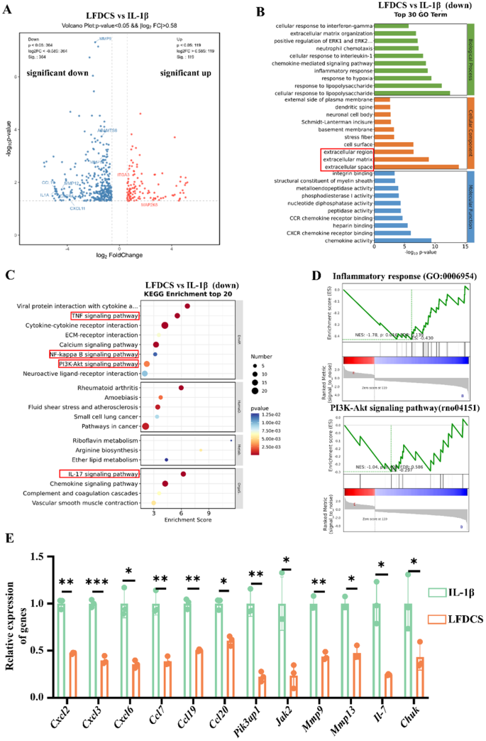
Subsequent, by analyzing the DEGs within the IL-1β group (chondrocytes handled with IL-1β) and the LFDCS group (chondrocytes handled with IL-1β and the leachate of LFDCS), 273 down-regulated genes and 73 up-regulated genes had been detected (Fig. S10B). The volcano plot outcomes confirmed down-regulation of pro-inflammatory genes (Ccl9, Cxcl11, and Il-1a), down-regulation of chondrocyte catabolic genes (Mmp12 and Mmp13), and up-regulation of ECM synthesis genes (Itga3 and Map2k6) within the LFDCS group compared to the IL-1β group (Fig. 10A). Gene ontology (GO) enrichment evaluation displayed that the results of LFDCS on the mobile parts of chondrocytes had been primarily centered on the regulation of extracellular areas, ECM, extracellular house, and ECM group, indicating that LFDCS ameliorated chondrocyte dysfunction by selling the ECM course of (Fig. 10B).
Subsequent evaluation by Kyoto Encyclopedia of Genes and Genomes (KEGG) displayed LFDCS regulates ECM synthesis by modulation of the TNF, NF-κB, IL-17, and PI3K/AKT signaling pathways (Fig. 10C). Moreover, we carried out gene set enrichment evaluation (GSEA) of the DEGs. The outcomes indicated that the LFDCS group down-regulated the chondrocyte inflammatory response-related and the PI3K/AKT signaling pathway in comparison with the IL-1β group (Fig. 10D). PI3K/AKT is a vital signaling pathway concerned in numerous capabilities that preserve mobile homeostasis [70]. Research present that down-regulation of the PI3K/AKT signaling pathway can alleviate chondrocyte deformation by activating mitochondrial autophagy [71, 72]. Concurrently, we verified the DEGs of curiosity within the RNA sequencing outcomes by qRT-PCR. These outcomes had been in accordance with the sequencing evaluation (Fig. 10E), which additional indicated that LFDCS improved chondrocyte metabolism by regulating the chondrocyte inflammation-associated and the PI3K/AKT signaling pathway.
As well as, chondrocyte operate was improved to a higher extent within the SerMA group (chondrocytes handled with IL-1β and the leachate of SerMA hydrogel microspheres) in comparison with the IL-1β group (Fig. 5 and 6). Furthermore, the LFDS exhibited a higher efficacy in enhancing cartilage operate than the SerMA did. We carried out KEGG enrichment evaluation of DEGs from the IL-1β and SerMA teams. The outcomes confirmed that the SerMA group inhibited chondrocyte inflammatory signaling pathway exercise, together with the NF-κB and chemokine signaling pathways (Fig. S10C). Furthermore, some research have demonstrated that SerMA might promote cartilage operate by way of the power of silk gum to fight IL-1β-induced chondrocyte dysfunction by scavenging ROS [73]. Our in vivo and in vitro research displayed that SerMA hydrogel microspheres, a bioactive materials, was additionally favorable to selling articular cartilage restore. Furthermore, inhibition of CD38 expression in chondrocytes ameliorates chondrocyte dysfunction and alleviates OA [29, 30].
Nevertheless, this research had some limitations. First, we didn’t discover the responsive launch of 78c@Lipo-FA in vivo. Second, we didn’t discover the mechanics of the LFDCS intimately. Third, the mechanism relating to the responsiveness of LFDCS to the native microenvironment of OA was not elucidated. Subsequently, follow-up experiments ought to deal with the characterization of the fabric and its particular mechanism of motion on chondrocytes and BMMs.
RNA sequencing of the IL-1β-treated chondrocytes with or with out LFDCS. . (A) DEGs between IL-1β and LFDCS teams from volcano map evaluation. (B) Mobile element of DEGs between IL-1β and LFDCS teams from GO enrichment evaluation. (C) Signaling pathways of DEGs between IL-1β and LFDCS teams from KEGG enrichment evaluation. (D) GSEA of inflammatory response and PI3K-Akt signaling pathway gene set in DEGs between IL-1β and LFDCS teams. (E) The expression of Cxcl2, Cxcl3, Cxcl6, Ccl7, Ccl19, Ccl20, Pik3ap1, Jak2, Mmp9, Mmp13, Il7, Chuk detected by qRT-PCR (n = 3). * p < 0.05, ** p < 0.01, *** p < 0.001. IL-1β: chondrocytes handled with IL-1β; LFDCS: chondrocytes handled with IL-1β and the leachate of LFDCS


