Characterization of N-CDs
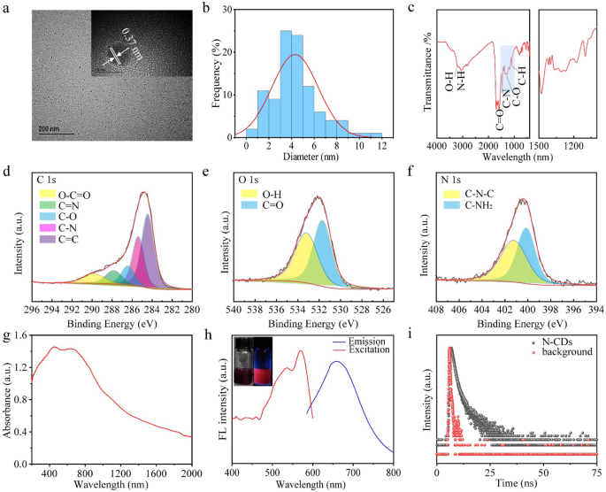
The transmission electron microscopy (TEM) picture of N-CDs (Fig. 2a) reveals spherical zero-dimensional constructions, with the high-resolution TEM picture (Fig. 2a inset) demonstrating a lattice construction with an interplanar spacing of about 0.37 nm. This spacing aligns with d-spacing of the graphitic carbon (002) lattice airplane [36]. Evaluation in Fig. 2b signifies that the common particle measurement of N-CDs is about 4.8 nm. The X-ray diffraction (XRD) sample (Fig. S1) reveals a definite peak at roughly 27°, comparable to the everyday (002) diffraction of graphitic carbon, indicative of a crystalline, layered construction just like graphene. The XRD information verify a well-defined graphitic construction, in line with earlier studies on N-doped CDs, the place graphitic nitrogen doping induces shifts in absorption and emission spectra [37]. Particularly, graphitic nitrogen narrows the vitality hole between the best occupied molecular orbital (HOMO) and the bottom unoccupied molecular orbital (LUMO), resulting in a crimson shift within the spectral response [12].
Within the Fourier remodel infrared (FT-IR) spectra (Fig. 2c), the absorption peaks at 3209 and 3080 cm− 1 for N-CDs correspond to O-H and N-H teams, respectively [37]. The height at 1580 cm− 1 is attributed to C = O absorption, whereas the height at 1477 cm− 1 arises from the uneven stretching vibration of C-N. Moreover, the absorption peak at 1309 cm− 1 is related to the stretching vibration of C-O [36].
The floor chemical properties of CDs had been additional investigated utilizing X-ray Photoelectron Spectroscopy (XPS). The complete spectrum (Fig. S2) revealed three main peaks at 289.7, 399.2, and 531.7 eV, comparable to the weather C, N, and O, respectively. The fundamental composition of N-CDs was calculated to be 74.66% of C, 11.23% of N and 14.11% of O, respectively (Desk S1). The high-resolution C1s spectrum (Fig. 2d) was deconvoluted into 5 peaks at 284.5, 285.6, 286.6, 287.9, and 289.8 eV, which correspond to C = C, C-N, C-O, C = N, and O-C-O purposeful teams, respectively [38, 39]. The O1s spectrum (Fig. 2e) displayed two peaks at 531.5 and 533.4 eV, attributed to O-H and C = O teams, respectively [40]. The N1s spectrum (Fig. 2f) revealed peaks at 400.2 eV and 401.5 eV, comparable to C-N-C and C-NH2, bonds, respectively, indicative of pyrrole nitrogen and graphitic nitrogen [9]. The XPS findings are in line with FT-IR spectroscopy outcomes, below high-pressure and high-temperature situations, the carboxyl teams of citric acid react with the amine teams of biuret by amide condensation, successfully incorporating nitrogen into CDs. Within the doped system, the introduction of graphitic nitrogen induces a crimson shift in seen mild absorption into the near-infrared spectral vary. This crimson shift is attributed to the presence of graphitic nitrogen, which narrows the H-L hole, with the extent of the shift rising with greater nitrogen doping concentrations [33]. Graphitic nitrogen doping creates mid-gap states inside the unique H-L hole of an undoped system, a results of extra electrons being donated into the unoccupied π* orbitals of the conjugated system [41]. The fluorescence properties of the graphitic nitrogen doped system exhibit a big narrowing of the H-L hole, resulting in a pronounced red-shifted emission in comparison with the undoped system [33]. The Raman spectra of N-CDs (Fig. S3) confirmed two broad peaks comparable to the D band and G band at about 1350 cm− 1 and 1615 cm− 1, respectively. The relative intensities of the D band to the G band (ID/IG = 0.95) indicated a excessive carbon lattice constructions within the N-CDs content material.
The optical properties of the N-CDs had been charaterized to judge their fluorescence efficiency. As proven within the UV-vis-NIR absorption spectrum (Fig. 2g), the N-CDs exhibited a broad absorption vary from 400 nm to 2000 nm, with sturdy absorption within the NIR-II area, indicating their distinctive NIR mild absorption capabilities. This superior NIR absorption is attributed to the excessive content material of graphitic nitrogen doping [33]. UV-vis-NIR spectrum of the N-CDs in answer (Fig. S4) additional corroborates their sturdy absorption within the NIR-II area. As proven in Fig. 2h, Underneath excitation at 565 nm, the N-CDs exhibited fluorescence emission with a peak at 660 nm (Fig. 2h), which lies inside the NIR-I organic window, enabling deeper fluorescence penetration. The fluorescence decay curve of N-CDs (Fig. 2i) revealed a fluorescence lifetime of 37.72 ns. Moreover, the emission depth of N-CDs assorted with the excitation wavelength (Fig. S5), demonstrating excitation-dependent emission habits, doubtless as a result of presence of a number of emission facilities on the floor of the N-CDs [27].
(a) Transmission Electron Microscopy (TEM) picture of N-CDs. (b) Particle measurement distribution curve for N-CDs. (c) Fourier Rework Infrared (FTIR) spectrum of N-CDs. (d–f) XPS spectra of N-CDs, C ingredient (d), O ingredient (e), N ingredient (f). (g) UV-vis-NIR spectrum of N-CDs. (h) Fluorescence emission spectrum of N-CDs. (i) Fluorescence decay curve of N-CDs in aqueous answer
Photothermal efficiency
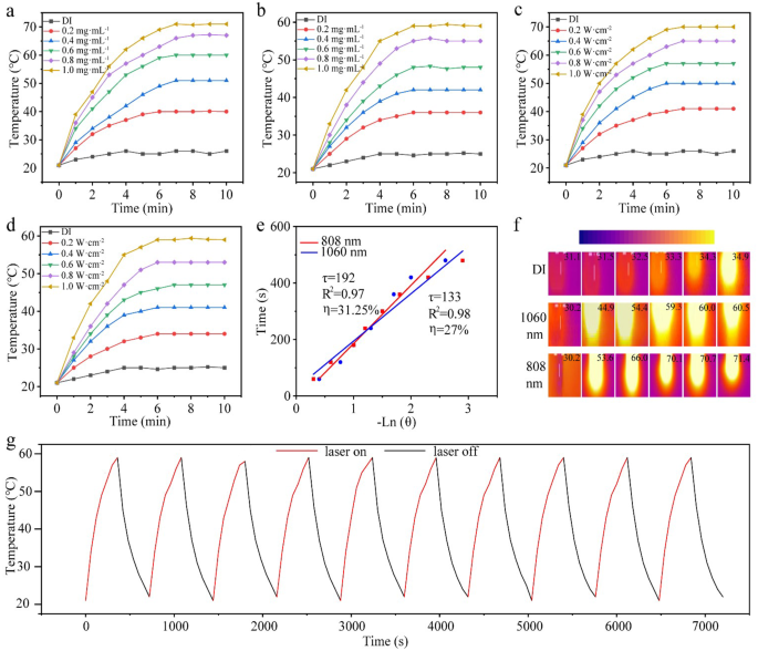
The UV-vis-NIR spectrum of the N-CDs revealed broad absorption peaks in each NIR-I and NIR-II areas, highlighting their potential as efficient near-infrared photothermal brokers. To judge their photothermal efficiency, laser sources at 808 nm and 1060 nm had been employed. The examine initially investigated the correlation between temperature rise and focus of N-CDs below 808 nm and 1060 nm laser irradiation (1.0 W·cm− 2). As proven in Fig. 3a, DI water displayed a minimal temperature improve of solely 5 ℃ below 808 nm irradiation. In distinction, a 0.2 mg·mL− 1 answer of N-CDs confirmed a temperature rise of 29 ℃, whereas options at 0.4 mg·mL− 1 and 0.6 mg·mL− 1 resulted in will increase of 34 ℃ and 38 ℃, respectively. The very best concentrations, 0.8 mg·mL− 1 and 1.0 mg·mL− 1, exhibited temperature rises of 40 ℃ and 45 ℃, respectively. The same pattern was noticed below 1060 nm laser irradiation (Fig. 3b), the place DI water elevated by solely 9 ℃, whereas the 0.2 mg·mL− 1 N-CDs answer rose by 24 ℃. Options at concentrations of 0.4, 0.6, 0.8, and 1.0 mg·mL− 1 demonstrated temperature rises of 28 ℃, 32 ℃, 34 ℃, and 38 ℃, respectively. These outcomes clearly exhibit the concentration-dependent photothermal impact of N-CDs.
The connection between the temperature rise of N-CDs (1.0 mg·mL− 1) and laser energy at 808 nm and 1060 nm was additionally examined. As proven in Fig. 3c, below 808 nm laser irradiation, the temperature will increase of 18 ℃, 21 ℃, 34 ℃, 38 ℃, and 43 ℃ had been noticed at energy densities of 0.2, 0.4, 0.6, 0.8, and 1.0 W·cm− 2, respectively. Equally, below 1060 nm laser irradiation (Fig. 3d), the temperature will increase of 13 ℃, 19 ℃, 26 ℃, 33 ℃, and 39 ℃ had been recorded on the similar energy densities. Moreover, primarily based on the cooling section information, the photothermal conversion effectivity of N-CDs was calculated to be 31.25% below 808 nm irradiation and 27.12% below 1060 nm irradiation (Fig. 3e). These values counsel that the N-CDs exhibit excessive photothermal conversion effectivity, making them extremely efficient as photothermal brokers for therapeutic purposes in tumor remedy below NIR mild.
Furthermore, Fig. 3f reveals the infrared thermal imaging of the N-CDs answer (1.0 mg·mL− 1) below each 808 nm (1.0 W·cm− 2) and 1060 nm (1.0 W·cm− 2) laser illumination. Vital temperature will increase had been noticed at every time interval (0, 2, 4, 6, 8, and 10 min), highlighting the distinctive photothermal effectivity of N-CDs. This effectivity might be attributed to a number of components. Firstly, the amide condensation response between carboxyl and amine teams leads to a nitrogen-rich floor, significantly the incorporation of graphitic nitrogen, which shifts the optical bandgap of N-CDs towards longer wavelengths [29]. Moreover, smaller nanoparticles exhibit greater light-to-heat conversion effectivity resulting from their elevated floor space and decreased mild scattering, minimizing vitality loss throughout irradiation. Lastly, the superb solubility and dispersibility of N-CDs in water facilitate speedy warmth switch to the encircling medium, additional enhancing temperature rise [9].
Moreover, the photothermal stability of N-CDs was assessed by ten heating and cooling cycles. As proven in Fig. 3g and S6, the heating-cooling cycle checks demonstrated that the N-CDs maintained constant photothermal efficiency throughout ten cycles below each 1060 nm and 808 nm illumination. No vital decline in photothermal efficiency was noticed, confirming the superb photothermal stability of N-CDs below NIR mild publicity.
Furthermore, the suspension stability of nanoparticles is decided by their Zeta potential. The Zeta potentials of N-CDs had been measured to be -16.01 mV in DI, -30.53 mV in PBS, and − 26.79 mV in RPMI-1640 medium (Fig. S7). Nanoparticles with a better floor cost, as indicated by Zeta potential, exhibit stronger repulsive pressure, resulting in secure dispersion and retention [42].
(a–d) Photothermal heating curves of N-CDs, displaying the impact of various concentrations (a–b), completely different powers (c–d). (e) Linear relationship between -lnθ and time. (f) Impact of heating of N-CDs below the thermal imaging digicam. (g) Ten photothermal cycles of N-CDs (1.0 mg·mL− 1) below 1060 nm laser illumination (1.0 W·cm− 2)
Antibacterial of N-CDs
Given their sturdy photothermal results in each the NIR-I and NIR-II areas, the near-infrared antibacterial capabilities of N-CDs had been evaluated utilizing Staphylococcus aureus as a mannequin organism. The antibacterial efficiency was examined at varied N-CDs concentrations (0.2, 0.4, 0.6, 0.8, and 1.0 mg·mL− 1) and completely different laser energy densities (0.2, 0.4 0.6, 0.8, and 1.0 W·cm− 2). As proven in Fig. S8 a and b, at mounted laser energy of 1.0 W·cm− 2 illumination, the variety of bacterial colonies dramatically decreased because the focus of the N-CDs answer elevated, reaching most antibacterial effectivity of 95% and 90% at 808 and 1060 nm, respectively. Moreover, the connection between antibacterial efficacy and laser energy was additionally studied. As depicted in Fig. S8 c and d, as the facility elevated to 1.0 W·cm− 2, the antibacterial effectivity reached 94% and 90% below 808 and 1060 nm irradiation, respectively. These outcomes verify that N-CDs successfully take in NIR mild and convert it into thermal vitality, producing adequate warmth to inhibit or remove Staphylococcus aureus.
Mobile experiments
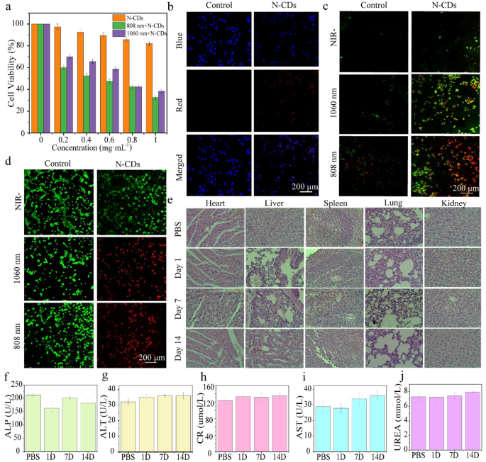
The PTT efficiency of N-CDs on the mobile degree was evaluated utilizing DU145 cells. Initially, the cytotoxicity of N-CDs was assessed utilizing the MTT assay [9]. To keep away from thermal injury to wholesome tissues, the laser energy density was maintained at 1.0 W·cm− 2 all through the experiment. Notably, cell viability remained excessive even at a N-CDs focus of 1.0 mg·mL− 1 with out laser irradiation, indicating low intrinsic cytotoxicity (Fig. 4a). Nonetheless, when subjected to 808–1060 nm lasers irradiation, cell viability decreased considerably. This implies that N-CDs exhibit appreciable phototoxicity below NIR mild, attributed to their glorious photothermal conversion potential, underscoring their potential for efficient photothermal remedy.
As well as, the mobile uptake potential of the N-CDs was evaluated utilizing laser confocal scanning microscopy. As depicted in Fig. 4b, the intensive overlap between the blue fluorescence (DAPI-stained nuclei) and the crimson fluorescence (internalized N-CDs) strongly means that the N-CDs had been effectively internalized by the cells reasonably than merely adhering to their floor. This environment friendly internalization additional highlights the numerous potential of N-CDs in tumor-targeted therapies.
Annexin V-FITC and Propidium Iodide (PI) staining had been utilized to evaluate cell apoptosis (Fig. 4c). Early apoptotic cells had been recognized by inexperienced fluorescence, whereas late apoptotic cells exhibited each inexperienced and crimson fluorescence. Within the management group, minimal apoptosis was noticed below all situations. In distinction, the experimental group handled with N-CDs confirmed a marked improve in apoptosis upon laser publicity, with most cells displaying twin inexperienced and crimson fluorescence below each 808 nm and 1060 nm illumination. These outcomes verify the numerous phototoxic results of N-CDs on cells below NIR mild publicity, highlighting their appreciable potential for photothermal remedy purposes.
The in vitro photothermal therapeutic efficacy of the N-CDs was additional validated utilizing a dual-staining experiment utilizing Acridine Orange (AO) and Propidium Iodide (PI) dyes. On this assay, stay cells had been stained inexperienced by AO, whereas lifeless cells appeared crimson resulting from PI staining. As illustrated in Fig. 4d, the management group, handled with PBS, exhibited predominantly inexperienced fluorescence, indicating minimal mobile injury. In distinction, cells handled with N-CDs and uncovered to the identical situations demonstrated pronounced crimson fluorescence at each 808 nm and 1060 nm wavelengths, indicting substantial cell demise. These findings align with the outcomes from the MTT assay, additional confirming efficient photothermal therapeutic potential of the N-CDs.
To additional assess the long-term in vivo biocompatibility of N-CDs, wholesome mice had been administered a dose of 5.0 mg·kg− 1. Histological evaluation of main organs (coronary heart, lungs, kidneys, liver, and spleen) was carried out utilizing H&E staining. The outcomes indicted no vital antagonistic results in mice handled with N-CDs answer over the primary 14 days (Fig. 4e). Moreover, liver purposeful markers, together with alanine aminotransferase (ALT), alkaline phosphatase (ALP), aspartate aminotransferase (AST), serum creatinine (CR), and urea (UREA) degree, confirmed no notable variations between the management and experimental teams (Fig. 4f-j).
Moreover, no vital modifications within the physique weight of the mice had been noticed in the course of the remedy interval (Fig. S9). The biocompatibility of N-CDs was additional supported by coagulation checks (Fig. S10). Mouse blood uncovered to PBS or the N-CDs answer maintained regular coagulation functionality, whereas publicity to water resulted in hemolysis and lack of coagulation potential, highlighting the secure interplay between N-CDs and organic programs. Taken collectively, these findings present compelling proof that N-CDs exhibit excessive biocompatibility, making them appropriate for PTT.
(a) Viability of DU145 cells incubated with options containing completely different concentrations of N-CDs below 808 nm and 1060 nm laser irradiation. (b) Uptake of N-CDs by DU145 cells incubated in PBS (management group) and N-CDs answer (experimental group). (c) Apoptosis imaging of DU145 cells incubated in PBS (management group) and N-CDs answer (experimental group) below darkish situations, with 1060 nm and 808 nm laser irradiation. (d) Stay/lifeless imaging of DU145 cells incubated in PBS (management group) and N-CDs answer (experimental group) below darkish situations, with 1060 nm and 808 nm laser irradiation. (e) H&E staining of main organs (coronary heart, lung, kidney, liver, and spleen) from wholesome mice 1, 7, and 14 days after intravenous injection of PBS and N-CDs. (f)-(j) Values of main purposeful components (alanine aminotransferase, ALT; alkaline phosphatase, ALP; aspartate aminotransferase, AST) within the liver, serum creatinine (CR), and urea (UREA) in wholesome mice 1, 7, and 14 days after intravenous injection of PBS and N-CDs
Animal experiments
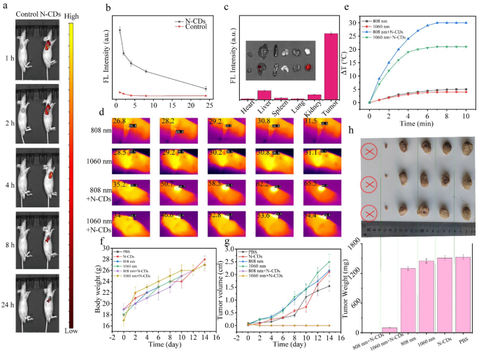
To evaluate the potential of N-CDs as a theranostic agent, their in vivo fluorescence imaging capabilities had been additional examined. Nude mice bearing subcutaneous DU145 tumors had been chosen because the animal mannequin, and N-CDs had been administered through intratumoral injection. As depicted in Fig. 5a and b, the fluorescence depth on the tumor website progressively elevated over time, peaking roughly 1 h post-injection earlier than regularly declining. This means that the uptake of N-CDs by tumor cells reaches its most inside 1 h. By 24 h, the fluorescence sign had almost disappeared, suggesting that the N-CDs had been extensively metabolized and cleared from the tumor website. Following intratumoral injection of N-CDs answer in mice, we carried out quantitative evaluation of fluorescence depth within the tumor tissue. As proven in Desk. S2, the fluorescence depth of N-CDs reached 9.092 × 10⁷ at 1 h post-injection, which subsequently decreased to 1.39 × 10⁷ after 24 h, yielding a ratio of roughly 0.153. This means an 84.7% discount of N-CDs on the tumor website inside 24 h, demonstrating substantial metabolic clearance. The dose of N-CDs was calculated to be injected into the mouse, leading to a complete mass of 10 µg. After 24 h, the metabolic price was 84.7%, thus, just one.53 µg of N-CDs remained inside the tumor of the mouse.
Moreover, to extra exactly assess the biodistribution of N-CDs, the mice had been humanely euthanized 24 h post-injection, and their main organs (coronary heart, liver, spleen, lung, and kidney) in addition to the tumor had been collected for additional evaluation. The fluorescence photos and quantified fluorescence depth values of the excised tissues (Fig. 5c) revealed minimal indicators in main organs, indicating environment friendly metabolism of N-CDs. Importantly, the fluorescence within the tissue samples was primarily inside the 500–600 nm vary. Notably, the crimson fluorescence emitted by N-CDs at 660 nm gives a big benefit. This particular crimson emission successfully reduces interference from intrinsic tissue autofluorescence, positioning N-CDs as a extremely promising candidate for stay imaging purposes.
Given the superb photothermal properties and excessive biocompatibility of N-CDs, their in vivo antitumor efficiency was assessed below NIR irradiation (Fig. S11). Tumor-bearing mice had been randomly divided into six teams and handled with PBS, N-CDs alone, 808 nm laser alone, 1060 nm laser alone, N-CDs + 808 nm laser, and N-CDs + 1060 nm laser, respectively. To reduce potential photodamage to wholesome tissue, the laser energy density was maintained at a comparatively low of 1.0 W·cm− 2. The infrared thermograms and temperature modifications on the tumor websites had been monitored utilizing an infrared digicam. As proven in Fig. 5d and e, the N-CDs + 808 nm and N-CDs + 1060 nm teams exhibited a big temperature improve on the tumor websites, whereas the teams receiving solely laser irradiation (808–1060 nm) confirmed minimal temperature modifications. After 4 min of irradiation, the tumor temperature within the N-CDs + 808 nm and N-CDs + 1060 nm teams rose from 35.2 °C to 58.5 °C and from 34.0 °C to 52.8 °C, respectively. In distinction, the 808 nm and 1060 nm management teams confirmed negligible temperature modifications (roughly 4.7 °C and a couple of.6 °C, respectively). As well as, no vital fluctuations in physique weight had been noticed in the course of the 14-day remedy interval (Fig. 5f), indicating that N-CDs cased minimal systemic unwanted side effects.
As proven in Fig. 5g and h, the tumors in N-CDs + 808 nm group had been utterly eradicated, whereas these within the N-CDs + 1060 nm group had been almost ablated. In distinction, tumors in management teams continued to develop quickly, visually confirming the superior tumor progress inhibition within the N-CDs + 808 nm and N-CDs + 1060 nm therapies. Moreover, no tumor recurrence was noticed in the course of the follow-up interval, demonstrating the effectiveness of N-CDs in stopping tumor regrowth.
Subsequently, histological evolution of main organs (coronary heart, lungs, kidneys, liver, and spleen) from the handled mice revealed no vital pathological modifications in both the management or experimental teams. This discovering additional helps the protection profile of N-CDs for vivo purposes (Fig. S12).
Moreover, histological examination of tumor tissue sections from the remedy teams revealed pronounced indicators of apoptosis and necrosis, whereas the management group exhibited no vital mobile injury (Fig. S13). These observations underscore the sturdy photothermal therapeutic efficacy of N-CDs, highlighting their potential as a promising and secure modality for most cancers remedy.
(a) Fluorescence visualization of DU145 tumor-bearing nude mice subsequent to the administration of the injection of N-CDs at corresponding time factors, with the crimson circle indicating the tumor space. (b) Fluorescence images of the main organs and tumor in DU145 tumor-bearing nude mice taken 24 h post-injection with N-CDs, together with the fluorescence intensities. (c) Fluorescence depth values on the tumor website of Nude mice with DU145 tumors at varied time intervals following the injection of N-CDs. (d) Infrared thermograms of the tumor websites in DU145 tumor-bearing nude mice from the management and experimental teams below 808–1060 nm (1.0 W·cm− 2) irradiation at completely different time intervals, and (e) the resultant temperature improve. (f) Weight change curves of tumor-bearing nude mice over 14 days below completely different therapies. (g) Tumor quantity change curves of tumor-bearing nude mice over 14 days below completely different therapies. (h) Schematic illustrations of tumors in several teams of tumor-bearing nude mice after 14 days


